4D1I

The structure of the GH35 beta-galactosidase Bgl35A from Cellvibrio japonicus


Experimental Data Snapshot

  • Method: X-RAY DIFFRACTION
  • Resolution: 1.80 Å
  • R-Value Free: 0.212 
  • R-Value Work: 0.185 
  • R-Value Observed: 0.186 

wwPDB Validation   3D Report Full Report


This is version 1.3 of the entry. See complete history


Literature

A Complex Gene Locus Enables Xyloglucan Utilization in the Model Saprophyte Cellvibrio Japonicus.

Larsbrink, J.Thompson, A.J.Lundqvist, M.Gardner, J.G.Davies, G.J.Brumer, H.

(2014) Mol Microbiol 94: 418

  • DOI: https://doi.org/10.1111/mmi.12776
  • Primary Citation of Related Structures:  
    4D1I, 4D1J

  • PubMed Abstract: 

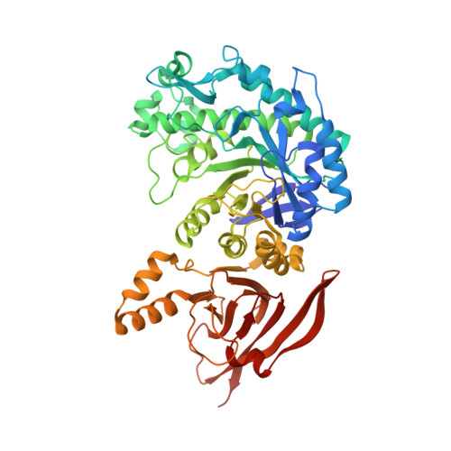
    The degradation of plant biomass by saprophytes is an ecologically important part of the global carbon cycle, which has also inspired a vast diversity of industrial enzyme applications. The xyloglucans (XyGs) constitute a family of ubiquitous and abundant plant cell wall polysaccharides, yet the enzymology of XyG saccharification is poorly studied. Here, we present the identification and molecular characterization of a complex genetic locus that is required for xyloglucan utilization by the model saprophyte Cellvibrio japonicus. In harness, transcriptomics, reverse genetics, enzyme kinetics, and structural biology indicate that the encoded cohort of an α-xylosidase, a β-galactosidase, and an α-l-fucosidase is specifically adapted for efficient, concerted saccharification of dicot (fucogalacto)xyloglucan oligosaccharides following import into the periplasm via an associated TonB-dependent receptor. The data support a biological model of xyloglucan degradation by C. japonicus with striking similarities - and notable differences - to the complex polysaccharide utilization loci of the Bacteroidetes.


  • Organizational Affiliation

    Division of Glycoscience, School of Biotechnology, Royal Institute of Technology (KTH), AlbaNova University Centre, 106 91, Stockholm, Sweden.


Macromolecules
Find similar proteins by:  (by identity cutoff)  |  3D Structure
Entity ID: 1
MoleculeChains Sequence LengthOrganismDetailsImage
BETA-GALACTOSIDASE, PUTATIVE, BGL35A
A, B, C, D, E
A, B, C, D, E, F, G, H
540Cellvibrio japonicus Ueda107Mutation(s): 0 
EC: 3.2.1.23
UniProt
Find proteins for B3PBE0 (Cellvibrio japonicus (strain Ueda107))
Explore B3PBE0 
Go to UniProtKB:  B3PBE0
Entity Groups  
Sequence Clusters30% Identity50% Identity70% Identity90% Identity95% Identity100% Identity
UniProt GroupB3PBE0
Sequence Annotations
Expand
  • Reference Sequence
Small Molecules
Ligands 2 Unique
IDChains Name / Formula / InChI Key2D Diagram3D Interactions
ACT
Query on ACT

Download Ideal Coordinates CCD File 
BA [auth H],
P [auth D],
Q [auth D]
ACETATE ION
C2 H3 O2
QTBSBXVTEAMEQO-UHFFFAOYSA-M
NA
Query on NA

Download Ideal Coordinates CCD File 
AA [auth H]
I [auth A]
J [auth B]
K [auth C]
L [auth C]
AA [auth H],
I [auth A],
J [auth B],
K [auth C],
L [auth C],
M [auth D],
N [auth D],
O [auth D],
R [auth E],
S [auth E],
T [auth E],
U [auth F],
V [auth F],
W [auth F],
X [auth G],
Y [auth G],
Z [auth G]
SODIUM ION
Na
FKNQFGJONOIPTF-UHFFFAOYSA-N
Experimental Data & Validation

Experimental Data

  • Method: X-RAY DIFFRACTION
  • Resolution: 1.80 Å
  • R-Value Free: 0.212 
  • R-Value Work: 0.185 
  • R-Value Observed: 0.186 
  • Space Group: P 1
Unit Cell:
Length ( Å )Angle ( ˚ )
a = 98.911α = 90.21
b = 115.783β = 90.25
c = 116.036γ = 90.38
Software Package:
Software NamePurpose
REFMACrefinement
XDSdata reduction
Aimlessdata scaling
PHASERphasing

Structure Validation

View Full Validation Report


Entry History 

Deposition Data

Revision History  (Full details and data files)

  • Version 1.0: 2014-05-28
    Type: Initial release
  • Version 1.1: 2014-10-22
    Changes: Database references
  • Version 1.2: 2020-03-11
    Changes: Derived calculations, Other
  • Version 1.3: 2023-12-20
    Changes: Data collection, Database references, Derived calculations, Refinement description