4D0T

GalNAc-T2 crystal soaked with UDP-GalNAc, EA2 peptide and manganese


Experimental Data Snapshot

  • Method: X-RAY DIFFRACTION
  • Resolution: 2.45 Å
  • R-Value Free: 0.241 
  • R-Value Work: 0.214 
  • R-Value Observed: 0.214 

wwPDB Validation   3D Report Full Report


Ligand Structure Quality Assessment 


This is version 1.4 of the entry. See complete history


Literature

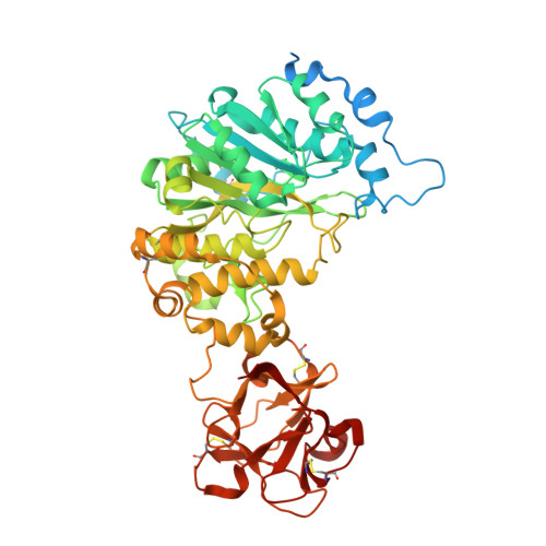
Substrate-Guided Front-Face Reaction Revealed by Combined Structural Snapshots and Metadynamics for the Polypeptide N-Acetylgalactosaminyltransferase 2.

Lira-Navarrete, E.Iglesias-Fernandez, J.Zandberg, W.F.Companon, I.Kong, Y.Corzana, F.Pinto, B.M.Clausen, H.Peregrina, J.M.Vocadlo, D.Rovira, C.Hurtado-Guerrero, R.

(2014) Angew Chem Int Ed Engl 53: 8206

  • DOI: https://doi.org/10.1002/anie.201402781
  • Primary Citation of Related Structures:  
    4D0T, 4D0Z, 4D11

  • PubMed Abstract: 

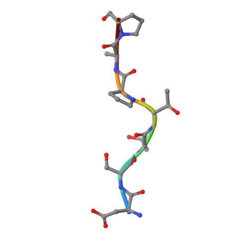
    The retaining glycosyltransferase GalNAc-T2 is a member of a large family of human polypeptide GalNAc-transferases that is responsible for the post-translational modification of many cell-surface proteins. By the use of combined structural and computational approaches, we provide the first set of structural snapshots of the enzyme during the catalytic cycle and combine these with quantum-mechanics/molecular-mechanics (QM/MM) metadynamics to unravel the catalytic mechanism of this retaining enzyme at the atomic-electronic level of detail. Our study provides a detailed structural rationale for an ordered bi-bi kinetic mechanism and reveals critical aspects of substrate recognition, which dictate the specificity for acceptor Thr versus Ser residues and enforce a front-face SN i-type reaction in which the substrate N-acetyl sugar substituent coordinates efficient glycosyl transfer.


  • Organizational Affiliation

    Institute of Biocomputation and Physics of Complex Systems (BIFI), University of Zaragoza, BIFI-IQFR (CSIC) Joint Unit, Mariano Esquillor s/n, Campus Rio Ebro, Edificio I+D, Fundacion ARAID, Edificio Pignatelli 36 (Spain).


Macromolecules
Find similar proteins by:  (by identity cutoff)  |  3D Structure
Entity ID: 1
MoleculeChains Sequence LengthOrganismDetailsImage
POLYPEPTIDE N-ACETYLGALACTOSAMINYLTRANSFERASE 2
A, B, C, D, E
A, B, C, D, E, F
571Homo sapiensMutation(s): 1 
EC: 2.4.1.41
UniProt & NIH Common Fund Data Resources
Find proteins for Q10471 (Homo sapiens)
Explore Q10471 
Go to UniProtKB:  Q10471
PHAROS:  Q10471
GTEx:  ENSG00000143641 
Entity Groups  
Sequence Clusters30% Identity50% Identity70% Identity90% Identity95% Identity100% Identity
UniProt GroupQ10471
Sequence Annotations
Expand
  • Reference Sequence

Find similar proteins by:  Sequence   |   3D Structure  

Entity ID: 2
MoleculeChains Sequence LengthOrganismDetailsImage
PEPTIDEG [auth P],
H [auth Z]
8synthetic constructMutation(s): 0 
Sequence Annotations
Expand
  • Reference Sequence
Small Molecules
Ligands 5 Unique
IDChains Name / Formula / InChI Key2D Diagram3D Interactions
UD2
Query on UD2

Download Ideal Coordinates CCD File 
DA [auth F],
L [auth A],
P [auth B],
V [auth D],
Z [auth E]
URIDINE-DIPHOSPHATE-N-ACETYLGALACTOSAMINE
C17 H27 N3 O17 P2
LFTYTUAZOPRMMI-NESSUJCYSA-N
UDP
Query on UDP

Download Ideal Coordinates CCD File 
R [auth C]URIDINE-5'-DIPHOSPHATE
C9 H14 N2 O12 P2
XCCTYIAWTASOJW-XVFCMESISA-N
NGA
Query on NGA

Download Ideal Coordinates CCD File 
EA [auth P]2-acetamido-2-deoxy-beta-D-galactopyranose
C8 H15 N O6
OVRNDRQMDRJTHS-JAJWTYFOSA-N
EDO
Query on EDO

Download Ideal Coordinates CCD File 
AA [auth E]
BA [auth E]
J [auth A]
K [auth A]
N [auth B]
AA [auth E],
BA [auth E],
J [auth A],
K [auth A],
N [auth B],
O [auth B],
S [auth C],
T [auth C],
W [auth D],
X [auth D]
1,2-ETHANEDIOL
C2 H6 O2
LYCAIKOWRPUZTN-UHFFFAOYSA-N
MN
Query on MN

Download Ideal Coordinates CCD File 
CA [auth F]
I [auth A]
M [auth B]
Q [auth C]
U [auth D]
CA [auth F],
I [auth A],
M [auth B],
Q [auth C],
U [auth D],
Y [auth E]
MANGANESE (II) ION
Mn
WAEMQWOKJMHJLA-UHFFFAOYSA-N
Experimental Data & Validation

Experimental Data

  • Method: X-RAY DIFFRACTION
  • Resolution: 2.45 Å
  • R-Value Free: 0.241 
  • R-Value Work: 0.214 
  • R-Value Observed: 0.214 
  • Space Group: P 21 21 21
Unit Cell:
Length ( Å )Angle ( ˚ )
a = 117.217α = 90
b = 122.372β = 90
c = 249.724γ = 90
Software Package:
Software NamePurpose
REFMACrefinement
XDSdata reduction
SCALEPACKdata scaling
PHASERphasing

Structure Validation

View Full Validation Report


Ligand Structure Quality Assessment 


Entry History 

Revision History  (Full details and data files)

  • Version 1.0: 2014-05-28
    Type: Initial release
  • Version 1.1: 2014-07-02
    Changes: Database references
  • Version 1.2: 2014-08-13
    Changes: Database references
  • Version 1.3: 2020-07-29
    Type: Remediation
    Reason: Carbohydrate remediation
    Changes: Advisory, Data collection, Derived calculations, Other, Structure summary
  • Version 1.4: 2023-12-20
    Changes: Data collection, Database references, Refinement description, Structure summary