4CUO

Banyan peroxidase with glycosylation


Experimental Data Snapshot

  • Method: X-RAY DIFFRACTION
  • Resolution: 1.67 Å
  • R-Value Free: 0.180 
  • R-Value Work: 0.157 
  • R-Value Observed: 0.158 

wwPDB Validation   3D Report Full Report


Ligand Structure Quality Assessment 


This is version 3.0 of the entry. See complete history


Literature

Post-Translational Modification and Extended Glycosylation Pattern of a Plant Latex Peroxidase of Native Source Characterized by X-Ray Crystallography.

Palm, G.J.Sharma, A.Kumari, M.Panjikar, S.Albrecht, D.Jagannadham, M.V.Hinrichs, W.

(2014) FEBS J 281: 4319

  • DOI: https://doi.org/10.1111/febs.12900
  • Primary Citation of Related Structures:  
    4CUO

  • PubMed Abstract: 

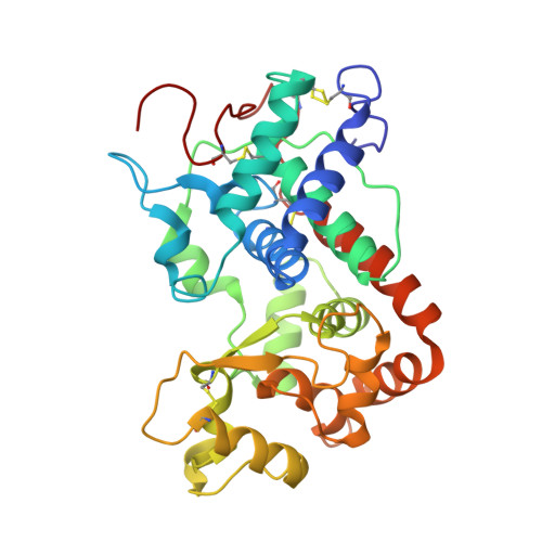
    The crystal structure of banyan peroxidase purified from the latex of Ficus benghalensis has been solved at 1.67 Å resolution by single-wavelength anomalous diffraction phasing. The refined structure includes 306 amino acid residues, a heme and two calcium ions. The protein belongs to class III peroxidases and is the first one from plant latex. Extensive glycosylation was observed with N-linked glycans attached to seven asparagine residues. The enzyme is stable with respect to a wide pH range, temperature, chemical denaturants and organic solvents, probably as a result of its high glycosylation. An unexpected post-translational modification of Asp290 was identified as succinimide moiety. Kinetic parameters of banyan peroxidase have been determined using various hydrogen donor substrates and hydrogen peroxide. Coordinates and structure factors have been deposited in the Protein Data Bank under accession number 4CUO.


  • Organizational Affiliation

    Institut für Biochemie, Universität Greifswald, Germany.


Macromolecules
Find similar proteins by:  (by identity cutoff)  |  3D Structure
Entity ID: 1
MoleculeChains Sequence LengthOrganismDetailsImage
BANYAN PEROXIDASE306Ficus benghalensisMutation(s): 0 
EC: 1.11.1.7
UniProt
Find proteins for A0A087WNH2 (Ficus benghalensis)
Explore A0A087WNH2 
Go to UniProtKB:  A0A087WNH2
Entity Groups  
Sequence Clusters30% Identity50% Identity70% Identity90% Identity95% Identity100% Identity
UniProt GroupA0A087WNH2
Sequence Annotations
Expand
  • Reference Sequence
Oligosaccharides

Help

Entity ID: 2
MoleculeChains Length2D Diagram Glycosylation3D Interactions
alpha-L-fucopyranose-(1-3)-2-acetamido-2-deoxy-beta-D-glucopyranose
B
2N-Glycosylation
Glycosylation Resources
GlyTouCan:  G80587NA
GlyCosmos:  G80587NA
GlyGen:  G80587NA
Entity ID: 3
MoleculeChains Length2D Diagram Glycosylation3D Interactions
alpha-D-xylopyranose-(1-2)-[alpha-D-mannopyranose-(1-3)][alpha-D-mannopyranose-(1-6)]beta-D-mannopyranose-(1-4)-2-acetamido-2-deoxy-beta-D-glucopyranose-(1-4)-[alpha-L-fucopyranose-(1-3)]2-acetamido-2-deoxy-beta-D-glucopyranose
C
7N-Glycosylation
Glycosylation Resources
GlyTouCan:  G98899DA
GlyCosmos:  G98899DA
GlyGen:  G98899DA
Entity ID: 4
MoleculeChains Length2D Diagram Glycosylation3D Interactions
alpha-L-fucopyranose-(1-3)-[2-acetamido-2-deoxy-beta-D-glucopyranose-(1-4)]2-acetamido-2-deoxy-beta-D-glucopyranose
D, E
3N-Glycosylation
Glycosylation Resources
GlyTouCan:  G11971MR
GlyCosmos:  G11971MR
GlyGen:  G11971MR
Small Molecules
Ligands 7 Unique
IDChains Name / Formula / InChI Key2D Diagram3D Interactions
HEM
Query on HEM

Download Ideal Coordinates CCD File 
F [auth A]PROTOPORPHYRIN IX CONTAINING FE
C34 H32 Fe N4 O4
KABFMIBPWCXCRK-RGGAHWMASA-L
NAG
Query on NAG

Download Ideal Coordinates CCD File 
G [auth A],
H [auth A],
I [auth A]
2-acetamido-2-deoxy-beta-D-glucopyranose
C8 H15 N O6
OVRNDRQMDRJTHS-FMDGEEDCSA-N
SO4
Query on SO4

Download Ideal Coordinates CCD File 
P [auth A],
Q [auth A],
R [auth A]
SULFATE ION
O4 S
QAOWNCQODCNURD-UHFFFAOYSA-L
CO3
Query on CO3

Download Ideal Coordinates CCD File 
S [auth A],
T [auth A],
U [auth A],
V [auth A]
CARBONATE ION
C O3
BVKZGUZCCUSVTD-UHFFFAOYSA-L
CA
Query on CA

Download Ideal Coordinates CCD File 
J [auth A],
K [auth A]
CALCIUM ION
Ca
BHPQYMZQTOCNFJ-UHFFFAOYSA-N
CL
Query on CL

Download Ideal Coordinates CCD File 
M [auth A],
N [auth A],
O [auth A]
CHLORIDE ION
Cl
VEXZGXHMUGYJMC-UHFFFAOYSA-M
NA
Query on NA

Download Ideal Coordinates CCD File 
L [auth A]SODIUM ION
Na
FKNQFGJONOIPTF-UHFFFAOYSA-N
Modified Residues  1 Unique
IDChains TypeFormula2D DiagramParent
SNN
Query on SNN
A
L-PEPTIDE LINKINGC4 H6 N2 O2ASN
Experimental Data & Validation

Experimental Data

  • Method: X-RAY DIFFRACTION
  • Resolution: 1.67 Å
  • R-Value Free: 0.180 
  • R-Value Work: 0.157 
  • R-Value Observed: 0.158 
  • Space Group: P 32 2 1
Unit Cell:
Length ( Å )Angle ( ˚ )
a = 73.115α = 90
b = 73.115β = 90
c = 164.596γ = 120
Software Package:
Software NamePurpose
REFMACrefinement
XDSdata reduction
XDSdata scaling
Auto-Rickshawphasing

Structure Validation

View Full Validation Report


Ligand Structure Quality Assessment 


Entry History 

Deposition Data

Revision History  (Full details and data files)

  • Version 1.0: 2014-07-23
    Type: Initial release
  • Version 1.1: 2014-10-01
    Changes: Database references
  • Version 1.2: 2019-07-17
    Changes: Data collection, Derived calculations
  • Version 2.0: 2020-07-29
    Type: Remediation
    Reason: Carbohydrate remediation
    Changes: Advisory, Atomic model, Data collection, Derived calculations, Other, Structure summary
  • Version 3.0: 2023-11-15
    Changes: Atomic model, Data collection, Database references, Derived calculations, Structure summary