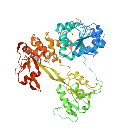
X-Ray Structure of the Pestivirus Ns3 Helicase and its Conformation in Solution.
Tortorici, M.A., Duquerroy, S., Kwok, J., Vonrhein, C., Perez, J., Lamp, B., Bricogne, G., Rumenapf, T., Vachette, P., Rey, F.A.(2015) J Virol 89: 4356
- PubMed: 25653438
- DOI: https://doi.org/10.1128/JVI.03165-14
- Primary Citation of Related Structures:
4CBG, 4CBH, 4CBI, 4CBL, 4CBM - PubMed Abstract:
Pestiviruses form a genus in the Flaviviridae family of small enveloped viruses with a positive-sense single-stranded RNA genome. Viral replication in this family requires the activity of a superfamily 2 RNA helicase contained in the C-terminal domain of nonstructural protein 3 (NS3). NS3 features two conserved RecA-like domains (D1 and D2) with ATPase activity, plus a third domain (D3) that is important for unwinding nucleic acid duplexes. We report here the X-ray structure of the pestivirus NS3 helicase domain (pNS3h) at a 2.5-Å resolution. The structure deviates significantly from that of NS3 of other genera in the Flaviviridae family in D3, as it contains two important insertions that result in a narrower nucleic acid binding groove. We also show that mutations in pNS3h that rescue viruses from which the core protein is deleted map to D3, suggesting that this domain may be involved in interactions that facilitate particle assembly. Finally, structural comparisons of the enzyme in different crystalline environments, together with the findings of small-angle X-ray-scattering studies in solution, show that D2 is mobile with respect to the rest of the enzyme, oscillating between closed and open conformations. Binding of a nonhydrolyzable ATP analog locks pNS3h in a conformation that is more compact than the closest apo-form in our crystals. Together, our results provide new insight and bring up new questions about pNS3h function during pestivirus replication.
Organizational Affiliation:
Unité de Virologie Structurale, Département de Virologie, Institut Pasteur, Paris, France CNRS, UMR 3569, Paris, France tortoric@pasteur.fr rey@pasteur.fr.