4C22

L-Fucose Isomerase In Complex With Fuculose


Experimental Data Snapshot

  • Method: X-RAY DIFFRACTION
  • Resolution: 2.70 Å
  • R-Value Free: 0.198 
  • R-Value Work: 0.144 
  • R-Value Observed: 0.147 

wwPDB Validation   3D Report Full Report


Ligand Structure Quality Assessment 


This is version 1.5 of the entry. See complete history


Literature

Structural and Functional Analysis of Fucose-Processing Enzymes from Streptococcus Pneumoniae.

Higgins, M.A.Suits, M.D.Marsters, C.Boraston, A.B.

(2014) J Mol Biol 426: 1469

  • DOI: https://doi.org/10.1016/j.jmb.2013.12.006
  • Primary Citation of Related Structures:  
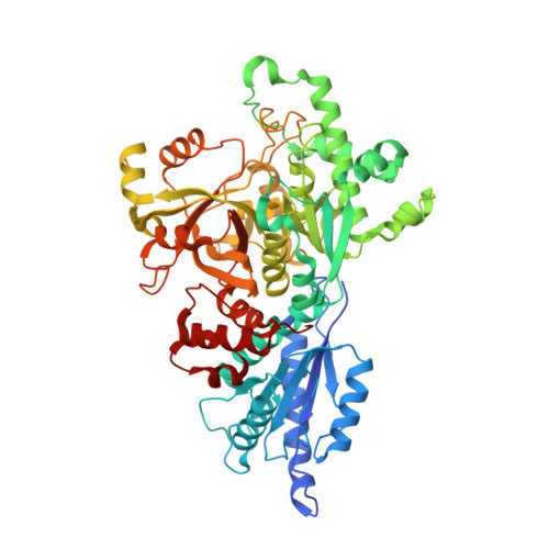
    4C20, 4C21, 4C22, 4C23, 4C24, 4C25

  • PubMed Abstract: 

    Fucose metabolism pathways are present in many bacterial species and typically contain the central fucose-processing enzymes fucose isomerase (FcsI), fuculose kinase (FcsK), and fuculose-1-phosphate aldolase (FcsA). Fucose initially undergoes isomerization by FcsI producing fuculose, which is then phosphorylated by FcsK. FcsA cleaves the fuculose-1-phosphate product into lactaldehyde and dihydroxyacetone phosphate, which can be incorporated into central metabolism allowing the bacterium to use fucose as an energy source. Streptococcus pneumoniae has fucose-processing operons containing homologs of FcsI, FcsK, and FcsA; however, this bacterium appears unable to utilize fucose as an energy source. To investigate this contradiction, we performed biochemical and structural studies of the S. pneumoniae fucose-processing enzymes SpFcsI, SpFcsK, and SpFcsA. These enzymes are demonstrated to act in a sequential manner to ultimately produce dihydroxyacetone phosphate and have structural features entirely consistent with their observed biochemical activities. Analogous to the regulation of the Escherichia coli fucose utilization operon, fuculose-1-phosphate appears to act as an inducing molecule for activation of the S. pneumoniae fucose operon. Despite our evidence that S. pneumoniae appears to have the appropriate regulatory and biochemical machinery for fucose metabolism, we confirmed the inability of the S. pneumoniae TIGR4 strain to grow on fucose or on the H-disaccharide, which is the probable substrate of the transporter for the pathway. On the basis of these observations, we postulate that the S. pneumoniae fucose-processing pathway has a non-metabolic role in the interaction of this bacterium with its human host.


  • Organizational Affiliation

    Department of Biochemistry and Microbiology, University of Victoria, PO Box 3055 STN CSC, Victoria, BC, V8W 3P6, Canada.


Macromolecules
Find similar proteins by:  (by identity cutoff)  |  3D Structure
Entity ID: 1
MoleculeChains Sequence LengthOrganismDetailsImage
L-FUCOSE ISOMERASE
A, B
604Streptococcus pneumoniae TIGR4Mutation(s): 0 
EC: 5.3.1.25
UniProt
Find proteins for Q97N97 (Streptococcus pneumoniae serotype 4 (strain ATCC BAA-334 / TIGR4))
Explore Q97N97 
Go to UniProtKB:  Q97N97
Entity Groups  
Sequence Clusters30% Identity50% Identity70% Identity90% Identity95% Identity100% Identity
UniProt GroupQ97N97
Sequence Annotations
Expand
  • Reference Sequence
Small Molecules
Ligands 4 Unique
IDChains Name / Formula / InChI Key2D Diagram3D Interactions
CVU
Query on CVU

Download Ideal Coordinates CCD File 
E [auth A],
O [auth B]
L-Fuculose open form
C6 H12 O5
QZNPNKJXABGCRC-LFRDXLMFSA-N
FUC
Query on FUC

Download Ideal Coordinates CCD File 
D [auth A],
N [auth B]
alpha-L-fucopyranose
C6 H12 O5
SHZGCJCMOBCMKK-SXUWKVJYSA-N
EDO
Query on EDO

Download Ideal Coordinates CCD File 
F [auth A]
G [auth A]
H [auth A]
I [auth A]
J [auth A]
F [auth A],
G [auth A],
H [auth A],
I [auth A],
J [auth A],
K [auth A],
L [auth A],
P [auth B],
Q [auth B],
R [auth B],
S [auth B],
T [auth B],
U [auth B],
V [auth B]
1,2-ETHANEDIOL
C2 H6 O2
LYCAIKOWRPUZTN-UHFFFAOYSA-N
MN
Query on MN

Download Ideal Coordinates CCD File 
C [auth A],
M [auth B]
MANGANESE (II) ION
Mn
WAEMQWOKJMHJLA-UHFFFAOYSA-N
Experimental Data & Validation

Experimental Data

  • Method: X-RAY DIFFRACTION
  • Resolution: 2.70 Å
  • R-Value Free: 0.198 
  • R-Value Work: 0.144 
  • R-Value Observed: 0.147 
  • Space Group: H 3 2
Unit Cell:
Length ( Å )Angle ( ˚ )
a = 151.45α = 90
b = 151.45β = 90
c = 406.4γ = 120
Software Package:
Software NamePurpose
REFMACrefinement
AutoProcessdata reduction
SCALAdata scaling
PHASERphasing

Structure Validation

View Full Validation Report


Ligand Structure Quality Assessment 


Entry History 

Deposition Data

Revision History  (Full details and data files)

  • Version 1.0: 2013-12-11
    Type: Initial release
  • Version 1.1: 2014-01-08
    Changes: Database references
  • Version 1.2: 2014-03-19
    Changes: Database references
  • Version 1.3: 2014-07-30
    Changes: Non-polymer description
  • Version 1.4: 2020-07-29
    Type: Remediation
    Reason: Carbohydrate remediation
    Changes: Data collection, Derived calculations, Other, Refinement description, Structure summary
  • Version 1.5: 2023-12-20
    Changes: Data collection, Database references, Derived calculations, Refinement description, Structure summary