4BT0

MuB is an AAAplus ATPase that forms helical filaments to control target selection for DNA transposition


Experimental Data Snapshot

  • Method: ELECTRON MICROSCOPY
  • Resolution: 17.0 Å
  • Aggregation State: FILAMENT 
  • Reconstruction Method: SINGLE PARTICLE 

wwPDB Validation   3D Report Full Report


This is version 1.3 of the entry. See complete history


Literature

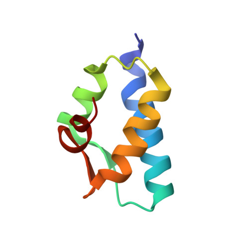
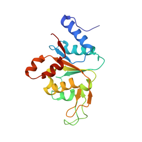
Mub is an Aaa+ ATPase that Forms Helical Filaments to Control Target Selection for DNA Transposition.

Mizuno, N.Dramicanin, M.Mizuuchi, M.Adam, J.Wang, Y.Han, Y.W.Yang, W.Steven, A.C.Mizuuchi, K.Ramon-Maiques, S.

(2013) Proc Natl Acad Sci U S A 110: E2441

  • DOI: https://doi.org/10.1073/pnas.1309499110
  • Primary Citation of Related Structures:  
    4BS1, 4BT0, 4BT1

  • PubMed Abstract: 

    MuB is an ATP-dependent nonspecific DNA-binding protein that regulates the activity of the MuA transposase and captures target DNA for transposition. Mechanistic understanding of MuB function has previously been hindered by MuB's poor solubility. Here we combine bioinformatic, mutagenic, biochemical, and electron microscopic analyses to unmask the structure and function of MuB. We demonstrate that MuB is an ATPase associated with diverse cellular activities (AAA+ ATPase) and forms ATP-dependent filaments with or without DNA. We also identify critical residues for MuB's ATPase, DNA binding, protein polymerization, and MuA interaction activities. Using single-particle electron microscopy, we show that MuB assembles into a helical filament, which binds the DNA in the axial channel. The helical parameters of the MuB filament do not match those of the coated DNA. Despite this protein-DNA symmetry mismatch, MuB does not deform the DNA duplex. These findings, together with the influence of MuB filament size on strand-transfer efficiency, lead to a model in which MuB-imposed symmetry transiently deforms the DNA at the boundary of the MuB filament and results in a bent DNA favored by MuA for transposition.


  • Organizational Affiliation

    Laboratory of Structural Biology, National Institute of Arthritis, Musculoskeletal, and Skin Diseases, National Institutes of Health, Bethesda, MD 20892, USA.


Macromolecules
Find similar proteins by:  (by identity cutoff)  |  3D Structure
Entity ID: 1
MoleculeChains Sequence LengthOrganismDetailsImage
TRANSCRIPTIONAL REGULATOR73Muvirus muMutation(s): 0 
UniProt
Find proteins for O67198 (Aquifex aeolicus (strain VF5))
Explore O67198 
Go to UniProtKB:  O67198
Entity Groups  
Sequence Clusters30% Identity50% Identity70% Identity90% Identity95% Identity100% Identity
UniProt GroupO67198
Sequence Annotations
Expand
  • Reference Sequence
Find similar proteins by:  (by identity cutoff)  |  3D Structure
Entity ID: 2
MoleculeChains Sequence LengthOrganismDetailsImage
TRANSCRIPTIONAL REGULATOR173Muvirus muMutation(s): 0 
UniProt
Find proteins for O67198 (Aquifex aeolicus (strain VF5))
Explore O67198 
Go to UniProtKB:  O67198
Entity Groups  
Sequence Clusters30% Identity50% Identity70% Identity90% Identity95% Identity100% Identity
UniProt GroupO67198
Sequence Annotations
Expand
  • Reference Sequence
Small Molecules
Experimental Data & Validation

Experimental Data

  • Method: ELECTRON MICROSCOPY
  • Resolution: 17.0 Å
  • Aggregation State: FILAMENT 
  • Reconstruction Method: SINGLE PARTICLE 
EM Software:
TaskSoftware PackageVersion
RECONSTRUCTIONBsoft
RECONSTRUCTIONEMAN
RECONSTRUCTIONSPIDER

Structure Validation

View Full Validation Report


Entry History 

Deposition Data

Revision History  (Full details and data files)

  • Version 1.0: 2013-07-03
    Type: Initial release
  • Version 1.1: 2013-07-17
    Changes: Database references
  • Version 1.2: 2013-08-07
    Changes: Derived calculations
  • Version 1.3: 2017-08-23
    Changes: Data collection