4BQL

Crystal structure of archaeal actin


Experimental Data Snapshot

  • Method: X-RAY DIFFRACTION
  • Resolution: 3.34 Å
  • R-Value Free: 0.248 
  • R-Value Work: 0.207 
  • R-Value Observed: 0.209 

wwPDB Validation   3D Report Full Report


Ligand Structure Quality Assessment 


This is version 1.1 of the entry. See complete history


Literature

Structure of Crenactin, an Archaeal Actin Homologue Active at 90Degc.

Lindas, A.C.Chruszcz, M.Bernander, R.Valegard, K.

(2014) Acta Crystallogr D Biol Crystallogr 70: 492

  • DOI: https://doi.org/10.1107/S1399004714000935
  • Primary Citation of Related Structures:  
    4BQL

  • PubMed Abstract: 

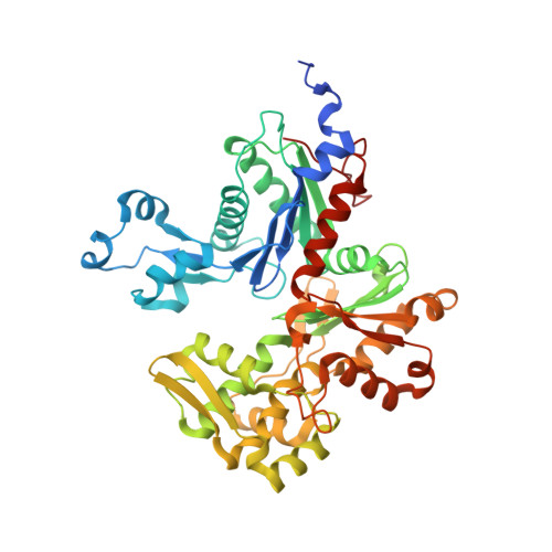
    The crystal structure of the archaeal actin, crenactin, from the rod-shaped hyperthermophilic (optimal growth at 90°C) crenarchaeon Pyrobaculum calidifontis is reported at 3.35 Å resolution. Despite low amino-acid sequence identity, the three-dimensional structure of the protein monomer is highly similar to those of eukaryotic actin and the bacterial MreB protein. Crenactin-specific features are also evident, as well as elements that are shared between crenactin and eukaryotic actin but are not found in MreB. In the crystal, crenactin monomers form right-handed helices, demonstrating that the protein is capable of forming filament-like structures. Monomer interactions in the helix, as well as interactions between crenactin and ADP in the nucleotide-binding pocket, are resolved at the atomic level and compared with those of actin and MreB. The results provide insights into the structural and functional properties of a heat-stable archaeal actin and contribute to the understanding of the evolution of actin-family proteins in the three domains of life.


  • Organizational Affiliation

    Department of Molecular Biosciences, The Wenner-Gren Institute, Stockholm University, Stockholm, Sweden.


Macromolecules
Find similar proteins by:  (by identity cutoff)  |  3D Structure
Entity ID: 1
MoleculeChains Sequence LengthOrganismDetailsImage
ACTIN/ACTIN FAMILY PROTEIN
A, B, C, D
455Pyrobaculum calidifontisMutation(s): 0 
UniProt
Find proteins for A3MWN5 (Pyrobaculum calidifontis (strain DSM 21063 / JCM 11548 / VA1))
Explore A3MWN5 
Go to UniProtKB:  A3MWN5
Entity Groups  
Sequence Clusters30% Identity50% Identity70% Identity90% Identity95% Identity100% Identity
UniProt GroupA3MWN5
Sequence Annotations
Expand
  • Reference Sequence
Experimental Data & Validation

Experimental Data

  • Method: X-RAY DIFFRACTION
  • Resolution: 3.34 Å
  • R-Value Free: 0.248 
  • R-Value Work: 0.207 
  • R-Value Observed: 0.209 
  • Space Group: P 21 21 21
Unit Cell:
Length ( Å )Angle ( ˚ )
a = 73.9α = 90
b = 88.217β = 90
c = 421.582γ = 90
Software Package:
Software NamePurpose
REFMACrefinement
HKL-3000data reduction
HKL-3000data scaling
HKL-3000phasing

Structure Validation

View Full Validation Report


Ligand Structure Quality Assessment 


Entry History 

Deposition Data

Revision History  (Full details and data files)

  • Version 1.0: 2014-02-12
    Type: Initial release
  • Version 1.1: 2014-02-26
    Changes: Database references