Exploring the Structural Basis of Substrate Preferences in Baeyer-Villiger Monooxygenases: Insight from Steroid Monooxygenase.
Franceschini, S., Van Beek, H.L., Pennetta, A., Martinoli, C., Fraaije, M.W., Mattevi, A.(2012) J Biol Chem 287: 22626
- PubMed: 22605340
- DOI: https://doi.org/10.1074/jbc.M112.372177
- Primary Citation of Related Structures:
4AOS, 4AOX, 4AP1, 4AP3 - PubMed Abstract:
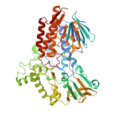
Steroid monooxygenase (STMO) from Rhodococcus rhodochrous catalyzes the Baeyer-Villiger conversion of progesterone into progesterone acetate using FAD as prosthetic group and NADPH as reducing cofactor. The enzyme shares high sequence similarity with well characterized Baeyer-Villiger monooxygenases, including phenylacetone monooxygenase and cyclohexanone monooxygenase. The comparative biochemical and structural analysis of STMO can be particularly insightful with regard to the understanding of the substrate-specificity properties of Baeyer-Villiger monooxygenases that are emerging as promising tools in biocatalytic applications and as targets for prodrug activation. The crystal structures of STMO in the native, NADP(+)-bound, and two mutant forms reveal structural details on this microbial steroid-degrading enzyme. The binding of the nicotinamide ring of NADP(+) is shifted with respect to the flavin compared with that observed in other monooxygenases of the same class. This finding fully supports the idea that NADP(H) adopts various positions during the catalytic cycle to perform its multiple functions in catalysis. The active site closely resembles that of phenylacetone monooxygenase. This observation led us to discover that STMO is capable of acting also on phenylacetone, which implies an impressive level of substrate promiscuity. The investigation of six mutants that target residues on the surface of the substrate-binding site reveals that enzymatic conversions of both progesterone and phenylacetone are largely insensitive to relatively drastic amino acid changes, with some mutants even displaying enhanced activity on progesterone. These features possibly reflect the fact that these enzymes are continuously evolving to acquire new activities, depending on the emerging availabilities of new compounds in the living environment.
Organizational Affiliation:
Department of Biology and Biotechnology, University of Pavia, Via Ferrata 9, 27100 Pavia, Italy.