Characterization of Staphylococcus Aureus Essb, an Integral Membrane Component of the Type Vii Secretion System: Atomic Resolution Crystal Structure of the Cytoplasmic Segment.
Zoltner, M., Fyfe, P.K., Palmer, T., Hunter, W.N.(2013) Biochem J 449: 469
- PubMed: 23098276
- DOI: https://doi.org/10.1042/BJ20121209
- Primary Citation of Related Structures:
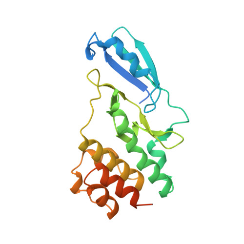
4ANN - PubMed Abstract:
The Type VII protein translocation/secretion system, unique to Gram-positive bacteria, is a key virulence determinant in Staphylococcus aureus. We aim to characterize the architecture of this secretion machinery and now describe the present study of S. aureus EssB, a 52 kDa bitopic membrane protein essential for secretion of the ESAT-6 (early secretory antigenic target of 6 kDa) family of proteins, the prototypic substrate of Type VII secretion. Full-length EssB was heterologously expressed in Escherichia coli, solubilized from the bacterial membrane, purified to homogeneity and shown to be dimeric. A C-terminal truncation, EssB∆C, and two soluble fragments termed EssB-N and EssB-C, predicted to occur on either side of the cytoplasmic membrane, have been successfully purified in a recombinant form, characterized and, together with the full-length protein, used in crystallization trials. EssB-N, the 25 kDa N-terminal cytoplasmic fragment, gave well-ordered crystals and we report the structure, determined by SAD (single-wavelength anomalous diffraction) targeting an SeMet (selenomethionine) derivative, refined to atomic (1.05 Å; 1 Å=0.1 nm) resolution. EssB-N is dimeric in solution, but crystallizes as a monomer and displays a fold comprised of two globular domains separated by a cleft. The structure is related to that of serine/threonine protein kinases and the present study identifies that the Type VII secretion system exploits and re-uses a stable modular entity and fold that has evolved to participate in protein-protein interactions in a similar fashion to the catalytically inert pseudokinases.
Organizational Affiliation:
College of Life Sciences, University of Dundee, Dow Street, Dundee DD1 5EH, UK.