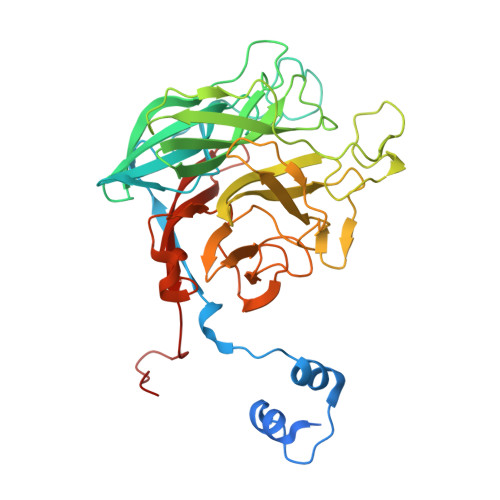
Analysis of Keystone Enzyme in Agar Hydrolysis Provides Insight Into the Degradation (of a Polysaccharide from) Red Seaweeds.
Hehemann, J.H., Smyth, L., Yadav, A., Vocadlo, D.J., Boraston, A.B.(2012) J Biol Chem 287: 13985
- PubMed: 22393053
- DOI: https://doi.org/10.1074/jbc.M112.345645
- Primary Citation of Related Structures:
4AK5, 4AK6, 4AK7 - PubMed Abstract:
Agars are abundant polysaccharides from marine red algae, and their chemical structure consists of alternating D-galactose and 3,6-anhydro-L-galactose residues, the latter of which are presumed to make the polymer recalcitrant to degradation by most terrestrial bacteria. Here we study a family 117 glycoside hydrolase (BpGH117) encoded within a recently discovered locus from the human gut bacterium Bacteroides plebeius. Consistent with this locus being involved in agarocolloid degradation, we show that BpGH117 is an exo-acting 3,6-anhydro-α-(1,3)-L-galactosidase that removes the 3,6-anhydrogalactose from the non-reducing end of neoagaro-oligosaccharides. A Michaelis complex of BpGH117 with neoagarobiose reveals the distortion of the constrained 3,6-anhydro-L-galactose into a conformation that favors catalysis. Furthermore, this complex, supported by analysis of site-directed mutants, provides evidence for an organization of the active site and positioning of the catalytic residues that are consistent with an inverting mechanism of catalysis and suggests that a histidine residue acts as the general acid. This latter feature differs from the vast majority of glycoside hydrolases, which use a carboxylic acid, highlighting the alternative strategies that enzymes may utilize in catalyzing the cleavage of glycosidic bonds.
Organizational Affiliation:
Department of Biochemistry and Microbiology, University of Victoria, PO Box 3055 STN CSC, Victoria, British Columbia V8W 3P6, Canada.