4AGS

Leishmania TDR1 - a unique trimeric glutathione transferase


Experimental Data Snapshot

  • Method: X-RAY DIFFRACTION
  • Resolution: 2.30 Å
  • R-Value Free: 0.208 
  • R-Value Work: 0.149 
  • R-Value Observed: 0.152 

wwPDB Validation   3D Report Full Report


Ligand Structure Quality Assessment 


This is version 1.2 of the entry. See complete history


Literature

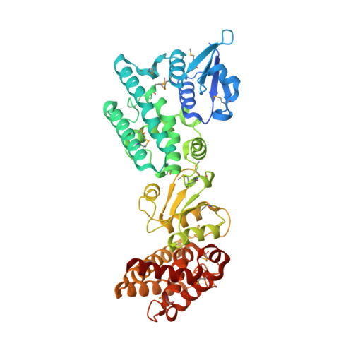
Leishmania Tdr1 Structure, a Unique Trimeric Glutathione Transferase Capable of Deglutathionylation and Antimonial Prodrug Activation.

Fyfe, P.K.Westrop, G.D.Silva, A.M.Coombs, G.H.Hunter, W.N.

(2012) Proc Natl Acad Sci U S A 109: 11693

  • DOI: https://doi.org/10.1073/pnas.1202593109
  • Primary Citation of Related Structures:  
    4AGS

  • PubMed Abstract: 

    Thiol-dependent reductase I (TDR1), an enzyme found in parasitic Leishmania species and Trypanosoma cruzi, is implicated in deglutathionylation and activation of antimonial prodrugs used to treat leishmaniasis. The 2.3 Å resolution structure of TDR1 reveals a unique trimer of subunits each containing two glutathione-S-transferase (GST) domains. The similarities of individual domains and comparisons with GST classes suggest that TDR1 evolved by gene duplication, diversification, and gene fusion; a combination of events previously unknown in the GST protein superfamily and potentially explaining the distinctive enzyme properties of TDR1. The deglutathionylation activity of TDR1 implies that glutathione itself has regulatory intracellular roles in addition to being a precursor for trypanothione, the major low mass thiol present in trypanosomatids. We propose that activation of antiparasite Sb(V)-drugs is a legacy of the deglutathionylation activity of TDR1 and involves processing glutathione adducts with concomitant reduction of the metalloid to active Sb(III) species.


  • Organizational Affiliation

    Division of Biological Chemistry and Drug Discovery, College of Life Sciences, University of Dundee, Dundee DD1 5EH, United Kingdom.


Macromolecules
Find similar proteins by:  (by identity cutoff)  |  3D Structure
Entity ID: 1
MoleculeChains Sequence LengthOrganismDetailsImage
THIOL-DEPENDENT REDUCTASE 1
A, B, C
471Leishmania infantumMutation(s): 0 
UniProt
Find proteins for A4I8P2 (Leishmania infantum)
Explore A4I8P2 
Go to UniProtKB:  A4I8P2
Entity Groups  
Sequence Clusters30% Identity50% Identity70% Identity90% Identity95% Identity100% Identity
UniProt GroupA4I8P2
Sequence Annotations
Expand
  • Reference Sequence
Small Molecules
Ligands 2 Unique
IDChains Name / Formula / InChI Key2D Diagram3D Interactions
GSH
Query on GSH

Download Ideal Coordinates CCD File 
D [auth A]
E [auth A]
L [auth B]
M [auth B]
S [auth C]
D [auth A],
E [auth A],
L [auth B],
M [auth B],
S [auth C],
T [auth C]
GLUTATHIONE
C10 H17 N3 O6 S
RWSXRVCMGQZWBV-WDSKDSINSA-N
EDO
Query on EDO

Download Ideal Coordinates CCD File 
F [auth A]
G [auth A]
H [auth A]
I [auth A]
J [auth A]
F [auth A],
G [auth A],
H [auth A],
I [auth A],
J [auth A],
K [auth A],
N [auth B],
O [auth B],
P [auth B],
Q [auth B],
R [auth B],
U [auth C],
V [auth C],
W [auth C],
X [auth C]
1,2-ETHANEDIOL
C2 H6 O2
LYCAIKOWRPUZTN-UHFFFAOYSA-N
Modified Residues  1 Unique
IDChains TypeFormula2D DiagramParent
MSE
Query on MSE
A, B, C
L-PEPTIDE LINKINGC5 H11 N O2 SeMET
Experimental Data & Validation

Experimental Data

  • Method: X-RAY DIFFRACTION
  • Resolution: 2.30 Å
  • R-Value Free: 0.208 
  • R-Value Work: 0.149 
  • R-Value Observed: 0.152 
  • Space Group: C 1 2 1
Unit Cell:
Length ( Å )Angle ( ˚ )
a = 197.477α = 90
b = 58.396β = 111.81
c = 160.385γ = 90
Software Package:
Software NamePurpose
REFMACrefinement
MOSFLMdata reduction
SCALAdata scaling
CRANKphasing

Structure Validation

View Full Validation Report


Ligand Structure Quality Assessment 


Entry History 

Deposition Data

Revision History  (Full details and data files)

  • Version 1.0: 2012-07-04
    Type: Initial release
  • Version 1.1: 2012-07-11
    Changes: Other
  • Version 1.2: 2012-08-01
    Changes: Database references