4A9C

Crystal structure of human SHIP2 in complex with biphenyl 2,3',4,5',6- pentakisphosphate


Experimental Data Snapshot

  • Method: X-RAY DIFFRACTION
  • Resolution: 2.10 Å
  • R-Value Free: 0.221 
  • R-Value Work: 0.201 
  • R-Value Observed: 0.202 

wwPDB Validation   3D Report Full Report


Ligand Structure Quality Assessment 


This is version 1.1 of the entry. See complete history


Literature

A Synthetic Polyphosphoinositide Headgroup Surrogate in Complex with Ship2 Provides a Rationale for Drug Discovery.

Mills, S.J.Persson, C.Cozier, G.Thomas, M.P.Tresaugues, L.Erneux, C.Riley, A.M.Nordlund, P.Potter, B.V.L.

(2012) ACS Chem Biol 7: 822

  • DOI: https://doi.org/10.1021/cb200494d
  • Primary Citation of Related Structures:  
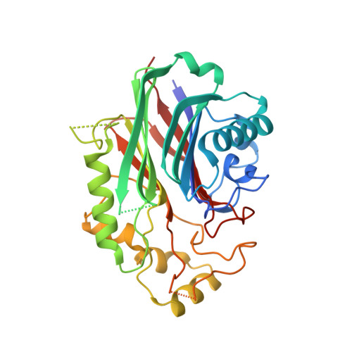
    4A9C

  • PubMed Abstract: 

    Phosphoinositides regulate many cellular processes, and cellular levels are controlled by kinases and phosphatases. SHIP2 (SH2 (Src homology 2)-domain-containing inositol-phosphatase-2) plays a critical role in phosphoinositide signaling, cleaving the 5-phosphate from phosphatidylinositol 3,4,5-trisphosphate. SHIP2 is thought to be involved in type-2 diabetes and obesity, conditions that could therefore be open to pharmacological modulation of the enzyme. However, rational design of SHIP2 inhibitors has been limited by the absence of a high-resolution structure. Here, we present a 2.1 Å resolution crystal structure of the phosphatase domain of SHIP2 bound to the synthetic ligand biphenyl 2,3',4,5',6-pentakisphosphate (BiPh(2,3',4,5',6)P(5)). BiPh(2,3',4,5',6)P(5) is not a SHIP2 substrate but inhibits Ins(1,3,4,5)P(4) hydrolysis with an IC(50) of 24.8 ± 3.0 μM, (K(m) for Ins(1,3,4,5)P(4) is 215 ± 28 μM). Molecular dynamics simulations suggest that when BiPh(2,3',4,5',6)P(5) binds to SHIP2, a flexible loop folds over and encloses the ligand. Compounds targeting such a closed conformation might therefore deliver SHIP2-specific drugs.


  • Organizational Affiliation

    Wolfson Laboratory of Medicinal Chemistry, Department of Pharmacy and Pharmacology, University of Bath, Bath BA2 7AY, U.K.


Macromolecules
Find similar proteins by:  (by identity cutoff)  |  3D Structure
Entity ID: 1
MoleculeChains Sequence LengthOrganismDetailsImage
PHOSPHATIDYLINOSITOL-3,4,5-TRISPHOSPHATE 5-PHOSPHATASE 2
A, B
316Homo sapiensMutation(s): 0 
EC: 3.1.3.86
UniProt & NIH Common Fund Data Resources
Find proteins for O15357 (Homo sapiens)
Explore O15357 
Go to UniProtKB:  O15357
PHAROS:  O15357
GTEx:  ENSG00000165458 
Entity Groups  
Sequence Clusters30% Identity50% Identity70% Identity90% Identity95% Identity100% Identity
UniProt GroupO15357
Sequence Annotations
Expand
  • Reference Sequence
Small Molecules
Ligands 1 Unique
IDChains Name / Formula / InChI Key2D Diagram3D Interactions
B5F
Query on B5F

Download Ideal Coordinates CCD File 
C [auth B]BIPHENYL 2,3',4,5',6-PENTAKISPHOSPHATE
C12 H15 O20 P5
ZCSNVIKBMFGCLE-UHFFFAOYSA-N
Binding Affinity Annotations 
IDSourceBinding Affinity
B5F Binding MOAD:  4A9C IC50: 2.48e+4 (nM) from 1 assay(s)
PDBBind:  4A9C IC50: 2.48e+4 (nM) from 1 assay(s)
Experimental Data & Validation

Experimental Data

  • Method: X-RAY DIFFRACTION
  • Resolution: 2.10 Å
  • R-Value Free: 0.221 
  • R-Value Work: 0.201 
  • R-Value Observed: 0.202 
  • Space Group: P 1 21 1
Unit Cell:
Length ( Å )Angle ( ˚ )
a = 45.246α = 90
b = 61.52β = 91.94
c = 114.742γ = 90
Software Package:
Software NamePurpose
BUSTERrefinement
XDSdata reduction
SCALAdata scaling
PHASERphasing

Structure Validation

View Full Validation Report


Ligand Structure Quality Assessment 


Entry History 

Revision History  (Full details and data files)

  • Version 1.0: 2012-05-30
    Type: Initial release
  • Version 1.1: 2023-12-20
    Changes: Data collection, Database references, Derived calculations, Other, Refinement description