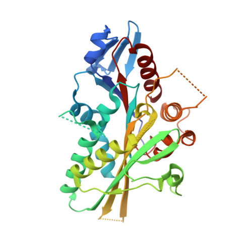
4A14

HUMAN KIF7, A KINESIN INVOLVED IN HEDGEHOG SIGNALLING


Experimental Data Snapshot

  • Method: X-RAY DIFFRACTION
  • Resolution: 1.60 Å
  • R-Value Free: 0.207 
  • R-Value Work: 0.182 
  • R-Value Observed: 0.184 

wwPDB Validation   3D Report Full Report


Ligand Structure Quality Assessment 


This is version 1.2 of the entry. See complete history


Literature

Structural Insights Into Human Kif7, a Kinesin Involved in Hedgehog Signalling.

Klejnot, M.Kozielski, F.

(2012) Acta Crystallogr D Biol Crystallogr 68: 154

  • DOI: https://doi.org/10.1107/S0907444911053042
  • Primary Citation of Related Structures:  
    2XT3, 4A14

  • PubMed Abstract: 

    Kif7, a member of the kinesin 4 superfamily, is implicated in a variety of diseases including Joubert, hydrolethalus and acrocallosal syndromes. It is also involved in primary cilium formation and the Hedgehog signalling pathway and may play a role in cancer. Its activity is crucial for embryonic development. Kif7 and Kif27, a closely related kinesin in the same subfamily, are orthologues of the Drosophila melanogaster kinesin-like protein Costal-2 (Cos2). In vertebrates, they work together to fulfil the role of the single Cos2 gene in Drosophila. Here, the high-resolution structure of the human Kif7 motor domain is reported and is compared with that of conventional kinesin, the founding member of the kinesin superfamily. These data are a first step towards structural characterization of a kinesin-4 family member and of this interesting molecular motor of medical significance.


  • Organizational Affiliation

    The Beatson Institute for Cancer Research, Garscube Estate, Switchback Road, Glasgow G61 1BD, Scotland. m.klejnot@beatson.gla.ac.uk


Macromolecules
Find similar proteins by:  (by identity cutoff)  |  3D Structure
Entity ID: 1
MoleculeChains Sequence LengthOrganismDetailsImage
KINESIN-LIKE PROTEIN KIF7344Homo sapiensMutation(s): 0 
UniProt & NIH Common Fund Data Resources
Find proteins for Q2M1P5 (Homo sapiens)
Explore Q2M1P5 
Go to UniProtKB:  Q2M1P5
PHAROS:  Q2M1P5
GTEx:  ENSG00000166813 
Entity Groups  
Sequence Clusters30% Identity50% Identity70% Identity90% Identity95% Identity100% Identity
UniProt GroupQ2M1P5
Sequence Annotations
Expand
  • Reference Sequence
Small Molecules
Ligands 2 Unique
IDChains Name / Formula / InChI Key2D Diagram3D Interactions
ADP
Query on ADP

Download Ideal Coordinates CCD File 
B [auth A]ADENOSINE-5'-DIPHOSPHATE
C10 H15 N5 O10 P2
XTWYTFMLZFPYCI-KQYNXXCUSA-N
MG
Query on MG

Download Ideal Coordinates CCD File 
C [auth A]MAGNESIUM ION
Mg
JLVVSXFLKOJNIY-UHFFFAOYSA-N
Experimental Data & Validation

Experimental Data

  • Method: X-RAY DIFFRACTION
  • Resolution: 1.60 Å
  • R-Value Free: 0.207 
  • R-Value Work: 0.182 
  • R-Value Observed: 0.184 
  • Space Group: P 21 21 21
Unit Cell:
Length ( Å )Angle ( ˚ )
a = 45.991α = 90
b = 79.799β = 90
c = 95.094γ = 90
Software Package:
Software NamePurpose
PHENIXrefinement
XDSdata reduction
SCALAdata scaling
MOLREPphasing

Structure Validation

View Full Validation Report


Ligand Structure Quality Assessment 


Entry History 

Deposition Data

Revision History  (Full details and data files)

  • Version 1.0: 2012-01-25
    Type: Initial release
  • Version 1.1: 2012-02-15
    Changes: Other
  • Version 1.2: 2023-12-20
    Changes: Data collection, Database references, Derived calculations, Other, Refinement description