Release of the cyano moiety in the crystal structure of N-cyanomethyl-N-(2-methoxyethyl)-daunomycin complexed with d(CGATCG).
Saminadin, P., Dautant, A., Mondon, M., Langlois D'estaintot, B., Courseille, C., Precigoux, G.(2000) Eur J Biochem 267: 457-464
- PubMed: 10632715
- DOI: https://doi.org/10.1046/j.1432-1327.2000.01017.x
- Primary Citation of Related Structures:
482D - PubMed Abstract:
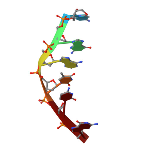
Doxorubicin is among the most widely used anthracycline in cancer chemotherapy. In an attempt to avoid the cardiotoxicity and drug resistance of doxorubicin therapy, several analogues were synthesized. The cyanomorpholinyl derivative is the most cytotoxic. They differ greatly from their parent compound in their biological and pharmacological properties, inducing cross-links in drug DNA complexes. The present study concerns N-cyanomethyl-N-(2-methoxyethyl)-daunomycin (CMDa), a synthetic analogue of cyanomorpholino-daunomycin. Compared to doxorubicin, CMDa displays a cytotoxic activity on L1210 leukemia cells at higher concentration but is effective on doxorubicin resistant cells. The results of fluorescence quenching experiments as well as the melting temperature (DeltaTm = 7.5 degrees C) studies are consistent with a drug molecule which intercalates between the DNA base pairs and stabilizes the DNA double helix. The crystal structure of CMDa complexed to the hexanucleotide d(CGATCG) has been determined at 1.5 A resolution. The complex crystallizes in the space group P41212 and is similar to other anthracycline-hexanucleotide complexes. In the crystal state, the observed densities indicate the formation of N-hydroxymethyl-N-(2-methoxyethyl)-daunomycin (HMDa) with the release of the cyano moiety without DNA alkylation. The formation of this degradation compound is discussed in relation with other drug modifications when binding to DNA. Comparison with two other drug-DNA crystal structures suggests a correlation between a slight change in DNA conformation and the nature of the amino sugar substituents at the N3' position located in the minor groove.
Organizational Affiliation:
Unité de Biophysique Structurale, CNRS UPRESA 5471, Université Bordeaux 1, Talence, France.