432D

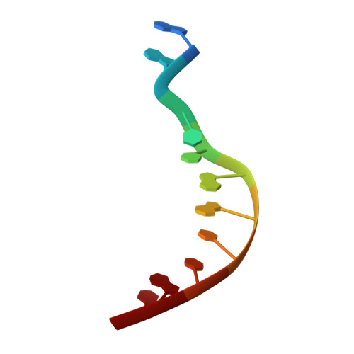
D(GGCCAATTGG) COMPLEXED WITH DAPI


Experimental Data Snapshot

  • Method: X-RAY DIFFRACTION
  • Resolution: 1.89 Å

wwPDB Validation   3D Report Full Report


Ligand Structure Quality Assessment 


This is version 1.5 of the entry. See complete history


Literature

Crystal structure of d(GGCCAATTGG) complexed with DAPI reveals novel binding mode.

Vlieghe, D.Sponer, J.Van Meervelt, L.

(1999) Biochemistry 38: 16443-16451

  • DOI: https://doi.org/10.1021/bi9907882
  • Primary Citation of Related Structures:  
    432D

  • PubMed Abstract: 

    The single-crystal X-ray structure of the complex between the minor groove binder 4',6-diamidino-2-phenylindole (DAPI) and d(GGCCAATTGG) reveals a novel way of off-centered binding, with an unique hydrogen bond between the minor groove binder and a CG base pair. Application of crystal engineering and cryocooling techniques helped to extend the resolution to 1.9 A, resulting in an unambiguous determination of drug conformation and orientation. The structure was refined to completion using SHELXL-93, resulting in a residual factor R of 18. 0% for 3562 reflections with F(o) > 4sigma(F(o)) including 81 water molecules. As the bulky NH(2)-group on guanine is believed to prevent drug binding in the minor groove, the nature and stability of the CG-DAPI contact was further addressed in full detail using ab initio quantum chemical methods. The amino groups involved in the guanine-drug interaction are substantially nonplanar, resulting in an energy gain of about 5 kcal/mol. The combined structural and theoretical data suggest that the guanine NH(2)-group does not destabilize the drug binding to an extent that it prevents complexation.


  • Organizational Affiliation

    Department of Chemistry, Katholieke Universiteit Leuven, Heverlee, Belgium.


Macromolecules

Find similar nucleic acids by:  Sequence   |   3D Structure  

Entity ID: 1
MoleculeChains LengthOrganismImage
DNA (5'-D(*GP*GP*CP*CP*AP*AP*TP*TP*GP*G)-3')
A, B
10N/A
Sequence Annotations
Expand
  • Reference Sequence
Small Molecules
Ligands 1 Unique
IDChains Name / Formula / InChI Key2D Diagram3D Interactions
DAP
Query on DAP

Download Ideal Coordinates CCD File 
C [auth A]6-AMIDINE-2-(4-AMIDINO-PHENYL)INDOLE
C16 H15 N5
FWBHETKCLVMNFS-UHFFFAOYSA-N
Experimental Data & Validation

Experimental Data

  • Method: X-RAY DIFFRACTION
  • Resolution: 1.89 Å
  • Space Group: P 21 21 21
Unit Cell:
Length ( Å )Angle ( ˚ )
a = 25.616α = 90
b = 36.563β = 90
c = 52.961γ = 90
Software Package:
Software NamePurpose
SHELXLrefinement
DENZOdata reduction
SCALEPACKdata scaling

Structure Validation

View Full Validation Report


Ligand Structure Quality Assessment 


Entry History 

Deposition Data

Revision History  (Full details and data files)

  • Version 1.0: 1999-12-18
    Type: Initial release
  • Version 1.1: 2008-04-26
    Changes: Version format compliance
  • Version 1.2: 2011-07-13
    Changes: Version format compliance
  • Version 1.3: 2017-10-04
    Changes: Refinement description
  • Version 1.4: 2023-12-27
    Changes: Data collection, Database references, Derived calculations
  • Version 1.5: 2024-04-03
    Changes: Refinement description