421P

THREE-DIMENSIONAL STRUCTURES OF H-RAS P21 MUTANTS: MOLECULAR BASIS FOR THEIR INABILITY TO FUNCTION AS SIGNAL SWITCH MOLECULES


Experimental Data Snapshot

  • Method: X-RAY DIFFRACTION
  • Resolution: 2.20 Å
  • R-Value Work: 0.192 
  • R-Value Observed: 0.192 

wwPDB Validation   3D Report Full Report


This is version 1.5 of the entry. See complete history


Literature

Three-dimensional structures of H-ras p21 mutants: molecular basis for their inability to function as signal switch molecules.

Krengel, U.Schlichting, I.Scherer, A.Schumann, R.Frech, M.John, J.Kabsch, W.Pai, E.F.Wittinghofer, A.

(1990) Cell 62: 539-548

  • DOI: https://doi.org/10.1016/0092-8674(90)90018-a
  • Primary Citation of Related Structures:  
    221P, 421P, 521P, 621P, 721P

  • PubMed Abstract: 

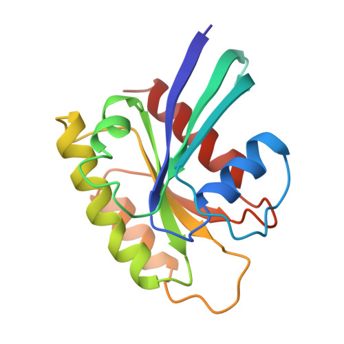
    The X-ray structures of the guanine nucleotide binding domains (amino acids 1-166) of five mutants of the H-ras oncogene product p21 were determined. The mutations described are Gly-12----Arg, Gly-12----Val, Gln-61----His, Gln-61----Leu, which are all oncogenic, and the effector region mutant Asp-38----Glu. The resolutions of the crystal structures range from 2.0 to 2.6 A. Cellular and mutant p21 proteins are almost identical, and the only significant differences are seen in loop L4 and in the vicinity of the gamma-phosphate. For the Gly-12 mutants the larger side chains interfere with GTP binding and/or hydrolysis. Gln-61 in cellular p21 adopts a conformation where it is able to catalyze GTP hydrolysis. This conformation has not been found for the mutants of Gln-61. Furthermore, Leu-61 cannot activate the nucleophilic water because of the chemical nature of its side chain. The D38E mutation preserves its ability to bind GAP.


  • Organizational Affiliation

    Max-Planck-Institut für medizinische Forschung Abteilung Biophysik, Heidelberg, Federal Republic of Germany.


Macromolecules
Find similar proteins by:  (by identity cutoff)  |  3D Structure
Entity ID: 1
MoleculeChains Sequence LengthOrganismDetailsImage
H-RAS P21 PROTEIN166Homo sapiensMutation(s): 1 
UniProt & NIH Common Fund Data Resources
Find proteins for P01112 (Homo sapiens)
Explore P01112 
Go to UniProtKB:  P01112
PHAROS:  P01112
GTEx:  ENSG00000174775 
Entity Groups  
Sequence Clusters30% Identity50% Identity70% Identity90% Identity95% Identity100% Identity
UniProt GroupP01112
Sequence Annotations
Expand
  • Reference Sequence
Small Molecules
Ligands 2 Unique
IDChains Name / Formula / InChI Key2D Diagram3D Interactions
GNP
Query on GNP

Download Ideal Coordinates CCD File 
C [auth A]PHOSPHOAMINOPHOSPHONIC ACID-GUANYLATE ESTER
C10 H17 N6 O13 P3
UQABYHGXWYXDTK-UUOKFMHZSA-N
MG
Query on MG

Download Ideal Coordinates CCD File 
B [auth A]MAGNESIUM ION
Mg
JLVVSXFLKOJNIY-UHFFFAOYSA-N
Experimental Data & Validation

Experimental Data

  • Method: X-RAY DIFFRACTION
  • Resolution: 2.20 Å
  • R-Value Work: 0.192 
  • R-Value Observed: 0.192 
  • Space Group: P 32 2 1
Unit Cell:
Length ( Å )Angle ( ˚ )
a = 40.4α = 90
b = 40.4β = 90
c = 162.2γ = 120
Software Package:
Software NamePurpose
X-PLORmodel building
X-PLORrefinement
X-PLORphasing

Structure Validation

View Full Validation Report


Entry History 

Deposition Data

Revision History  (Full details and data files)

  • Version 1.0: 1994-01-31
    Type: Initial release
  • Version 1.1: 2008-03-03
    Changes: Version format compliance
  • Version 1.2: 2011-07-13
    Changes: Version format compliance
  • Version 1.3: 2012-03-07
    Changes: Database references
  • Version 1.4: 2017-11-29
    Changes: Derived calculations, Other
  • Version 1.5: 2024-02-28
    Changes: Data collection, Database references, Derived calculations