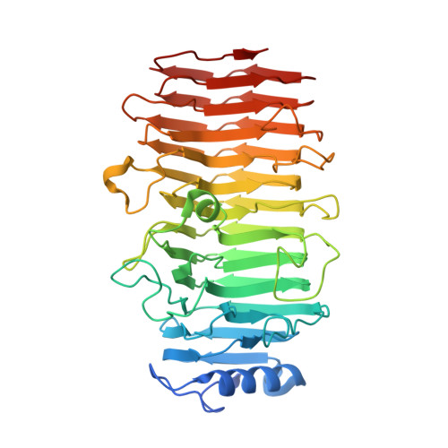
Structure of the Streptococcus Pneumoniae Surface Protein and Adhesin Pfba.
Suits, M.D., Boraston, A.B.(2013) PLoS One 8: 67190
- PubMed: 23894284
- DOI: https://doi.org/10.1371/journal.pone.0067190
- Primary Citation of Related Structures:
3ZPP - PubMed Abstract:
PfbA (plasmin- and fibronectin-binding protein A) is an extracellular Streptococcus pneumoniae cell-wall attached surface protein that binds to fibronectin, plasmin, and plasminogen. Here we present a structural analysis of the surface exposed domains of PfbA using a combined approach of X-ray crystallography and small-angle X-ray scattering (SAXS). The crystal structure of the PfbA core domain, here called PfbAβ, determined to 2.28 Å resolution revealed an elongated 12-stranded parallel β-helix fold, which structure-based comparisons reveal is most similar to proteins with carbohydrate modifying activity. A notable feature of the PfbAβ is an extensive cleft on one face of the protein with electrochemical and spatial features that are analogous to structurally similar carbohydrate-active enzymes utilizing this feature for substrate accommodation. Though this cleft displays a combination of basic amino acid residues and solvent exposed aromatic amino acids that are distinct features for recognition of carbohydrates, no obvious arrangement of amino acid side chains that would constitute catalytic machinery is evident. The pseudo-atomic SAXS model of a larger fragment of PfbA suggests that it has a relatively well-ordered structure with the N-terminal and core domains of PfbA adopting an extend organization and reveals a novel structural class of surface exposed pneumococcal matrix molecule adhesins.
Organizational Affiliation:
Department of Biochemistry and Microbiology, University of Victoria, Victoria, British Columbia, Canada.