3ZNM

H5 Haemagglutinin in Complex with Sialyl-Lewis X


Experimental Data Snapshot

  • Method: X-RAY DIFFRACTION
  • Resolution: 2.40 Å
  • R-Value Free: 0.256 
  • R-Value Work: 0.238 
  • R-Value Observed: 0.239 

wwPDB Validation   3D Report Full Report


Ligand Structure Quality Assessment 


This is version 2.1 of the entry. See complete history


Literature

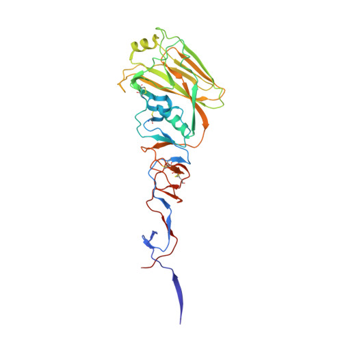
Recognition of Sulphated and Fucosylated Receptor Sialosides by A/Vietnam/1194/2004 (H5N1) Influenza Virus.

Xiong, X.Tuzikov, A.Coombs, P.Martin, S.Walker, P.A.Gamblin, S.J.Bovin, N.Skehel, J.J.

(2013) Virus Res 178: 12


Macromolecules
Find similar proteins by:  (by identity cutoff)  |  3D Structure
Entity ID: 1
MoleculeChains Sequence LengthOrganismDetailsImage
HAEMAGGLUTININ
A, C, E
326Influenza A virus (A/Vietnam/1194/2004(H5N1))Mutation(s): 0 
UniProt
Find proteins for Q6DQ34 (Influenza A virus)
Explore Q6DQ34 
Go to UniProtKB:  Q6DQ34
Entity Groups  
Sequence Clusters30% Identity50% Identity70% Identity90% Identity95% Identity100% Identity
UniProt GroupQ6DQ34
Sequence Annotations
Expand
  • Reference Sequence
Find similar proteins by:  (by identity cutoff)  |  3D Structure
Entity ID: 2
MoleculeChains Sequence LengthOrganismDetailsImage
HAEMAGGLUTININ
B, D, F
166Influenza A virus (A/Vietnam/1194/2004(H5N1))Mutation(s): 0 
UniProt
Find proteins for Q6DQ34 (Influenza A virus)
Explore Q6DQ34 
Go to UniProtKB:  Q6DQ34
Entity Groups  
Sequence Clusters30% Identity50% Identity70% Identity90% Identity95% Identity100% Identity
UniProt GroupQ6DQ34
Sequence Annotations
Expand
  • Reference Sequence
Oligosaccharides

Help

Entity ID: 3
MoleculeChains Length2D Diagram Glycosylation3D Interactions
N-acetyl-alpha-neuraminic acid-(2-3)-alpha-D-galactopyranose-(1-4)-[alpha-L-fucopyranose-(1-3)]2-acetamido-2-deoxy-beta-D-glucopyranose
G, H, I
4N/A
Glycosylation Resources
GlyTouCan:  G85849LM
GlyCosmos:  G85849LM
GlyGen:  G85849LM
Small Molecules
Ligands 2 Unique
IDChains Name / Formula / InChI Key2D Diagram3D Interactions
EPE
Query on EPE

Download Ideal Coordinates CCD File 
M [auth B],
Q [auth D],
U [auth F]
4-(2-HYDROXYETHYL)-1-PIPERAZINE ETHANESULFONIC ACID
C8 H18 N2 O4 S
JKMHFZQWWAIEOD-UHFFFAOYSA-N
NAG
Query on NAG

Download Ideal Coordinates CCD File 
J [auth A]
K [auth A]
L [auth B]
N [auth C]
O [auth C]
J [auth A],
K [auth A],
L [auth B],
N [auth C],
O [auth C],
P [auth D],
R [auth E],
S [auth E],
T [auth F]
2-acetamido-2-deoxy-beta-D-glucopyranose
C8 H15 N O6
OVRNDRQMDRJTHS-FMDGEEDCSA-N
Experimental Data & Validation

Experimental Data

  • Method: X-RAY DIFFRACTION
  • Resolution: 2.40 Å
  • R-Value Free: 0.256 
  • R-Value Work: 0.238 
  • R-Value Observed: 0.239 
  • Space Group: C 1 2 1
Unit Cell:
Length ( Å )Angle ( ˚ )
a = 175.56α = 90
b = 101.36β = 111.29
c = 161.2γ = 90
Software Package:
Software NamePurpose
REFMACrefinement
MOSFLMdata reduction
SCALAdata scaling
PHASERphasing

Structure Validation

View Full Validation Report


Ligand Structure Quality Assessment 


Entry History 

Deposition Data

Revision History  (Full details and data files)

  • Version 1.0: 2013-09-25
    Type: Initial release
  • Version 1.1: 2013-11-27
    Changes: Database references
  • Version 1.2: 2015-01-21
    Changes: Atomic model, Derived calculations, Non-polymer description, Other, Refinement description
  • Version 2.0: 2020-07-29
    Type: Remediation
    Reason: Carbohydrate remediation
    Changes: Advisory, Atomic model, Data collection, Derived calculations, Other, Structure summary
  • Version 2.1: 2023-12-20
    Changes: Data collection, Database references, Refinement description, Structure summary