3ZLW

Crystal structure of MEK1 in complex with fragment 3


Experimental Data Snapshot

  • Method: X-RAY DIFFRACTION
  • Resolution: 2.12 Å
  • R-Value Free: 0.248 
  • R-Value Work: 0.196 
  • R-Value Observed: 0.198 

wwPDB Validation   3D Report Full Report


Ligand Structure Quality Assessment 


This is version 2.0 of the entry. See complete history


Literature

The Use of Virtual Screening and Differential Scanning Fluorimetry for the Rapid Identification of Fragments Active Against Mek1.

Amaning, K.Lowinski, M.Vallee, F.Steier, V.Marcireau, C.Ugolini, A.Delorme, C.Foucalt, F.Mccort, G.Derimay, N.Andouche, C.Vougier, S.Llopart, S.Halland, N.Rak, A.

(2013) Bioorg Med Chem Lett 23: 3620

  • DOI: https://doi.org/10.1016/j.bmcl.2013.04.003
  • Primary Citation of Related Structures:  
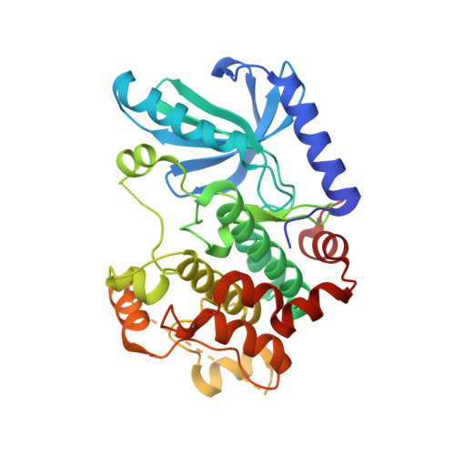
    3ZLS, 3ZLW, 3ZLX, 3ZLY, 3ZM4

  • PubMed Abstract: 

    We report the analysis of an in-house fragment screening campaign for the oncology target MEK1. The application of virtual screening (VS) as a primary fragment screening approach, followed by biophysical validation using differential screening fluorimetry (DSF), with resultant binding mode determination by X-ray crystallography (X-ray), is presented as the most time and cost-effective combination of in silico and in vitro methods to identify fragments. We demonstrate the effectiveness of the VS-DSF workflow for the early identification of fragments to both 'jump-start' the drug discovery project and to complement biochemical screening data.


  • Organizational Affiliation

    Sanofi, Structure Design and Informatics,13 Quai Guesde, B.P.14, F-94408 Vitry-Sur-Seine, France. kwame.amaning@sanofi.com


Macromolecules
Find similar proteins by:  (by identity cutoff)  |  3D Structure
Entity ID: 1
MoleculeChains Sequence LengthOrganismDetailsImage
DUAL SPECIFICITY MITOGEN-ACTIVATED PROTEIN KINASE KINASE 1 MAPK/ERK KINASE 1, MEK 1, MEK1348Homo sapiensMutation(s): 3 
EC: 2.7.12.2
UniProt & NIH Common Fund Data Resources
Find proteins for Q02750 (Homo sapiens)
Explore Q02750 
Go to UniProtKB:  Q02750
PHAROS:  Q02750
GTEx:  ENSG00000169032 
Entity Groups  
Sequence Clusters30% Identity50% Identity70% Identity90% Identity95% Identity100% Identity
UniProt GroupQ02750
Sequence Annotations
Expand
  • Reference Sequence
Small Molecules
Ligands 1 Unique
IDChains Name / Formula / InChI Key2D Diagram3D Interactions
MT8
Query on MT8

Download Ideal Coordinates CCD File 
B [auth A](1R)-1-hydroxy-1-methyl-2,3,6,7-tetrahydro-1H,5H-pyrido[3,2,1-ij]quinolin-5-one
C13 H15 N O2
FHMNDLFMEHZYLO-CYBMUJFWSA-N
Binding Affinity Annotations 
IDSourceBinding Affinity
MT8 Binding MOAD:  3ZLW IC50: 1.98e+5 (nM) from 1 assay(s)
BindingDB:  3ZLW IC50: 1.81e+4 (nM) from 1 assay(s)
PDBBind:  3ZLW IC50: 1.98e+5 (nM) from 1 assay(s)
Experimental Data & Validation

Experimental Data

  • Method: X-RAY DIFFRACTION
  • Resolution: 2.12 Å
  • R-Value Free: 0.248 
  • R-Value Work: 0.196 
  • R-Value Observed: 0.198 
  • Space Group: P 61 2 2
Unit Cell:
Length ( Å )Angle ( ˚ )
a = 77.059α = 90
b = 77.059β = 90
c = 221.8γ = 120
Software Package:
Software NamePurpose
BUSTERrefinement
PHASERphasing

Structure Validation

View Full Validation Report


Ligand Structure Quality Assessment 


Entry History 

Revision History  (Full details and data files)

  • Version 1.0: 2013-05-22
    Type: Initial release
  • Version 1.1: 2013-05-29
    Changes: Database references
  • Version 1.2: 2018-05-02
    Changes: Data collection
  • Version 2.0: 2019-04-24
    Changes: Atomic model, Data collection, Source and taxonomy