Single Mutation Alters the Substrate Specificity of l-Amino Acid Ligase
Tsuda, T., Asami, M., Koguchi, Y., Kojima, S.(2014) Biochemistry 53: 2650-2660
- PubMed: 24702628
- DOI: https://doi.org/10.1021/bi500292b
- Primary Citation of Related Structures:
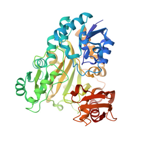
3WNZ, 3WO0, 3WO1 - PubMed Abstract:
L-Amino acid ligase (Lal) catalyzes the formation of a dipeptide from two L-amino acids in an ATP-dependent manner and belongs to the ATP-grasp superfamily. Bacillus subtilis YwfE, the first identified Lal, produces the dipeptide antibiotic bacilysin, which consists of L-Ala and L-anticapsin. Its substrate specificity is restricted to smaller amino acids such as L-Ala for the N-terminal end of the dipeptide, whereas a wide range of hydrophobic amino acids including L-Phe and L-Met are recognized for the C-terminal end in vitro. We determined the crystal structures of YwfE with bound ADP-Mg(2+)-Pi and ADP-Mg(2+)-L-Ala at 1.9 and 2.0 Å resolutions, respectively. On the basis of these structures, we generated point mutants of residues that are considered to participate in the recognition of L-Ala and measured their ATPase activity. The conserved Arg328 is suggested to be a crucial residue for L-Ala recognition and catalysis. The mutation of Trp332 to Ala caused the enzyme to hydrolyze ATP, even in the absence of l-Ala, and the structure of this mutant protein appeared to show a cavity in the N-terminal substrate-binding pocket. These results suggest that Trp332 plays a key role in restricting the substrate specificity to smaller amino acids such as L-Ala. Moreover, Trp332 mutants can alter the substrate specificity and activity depending on the size and shape of substituted amino acids. These observations provide sufficient scope for the rational design of Lal to produce desirable dipeptides. We propose that the positioning of the conserved Arg residue in Lal is important for enantioselective recognition of L-amino acids.
Organizational Affiliation:
Department of Life Science, Faculty of Science, Gakushuin University , 1-5-1 Mejiro, Toshima-ku, Tokyo 171-8588, Japan.