3WG2

Crystal structure of Agrocybe cylindracea galectin mutant (N46A)


Experimental Data Snapshot

  • Method: X-RAY DIFFRACTION
  • Resolution: 2.20 Å
  • R-Value Free: 0.244 
  • R-Value Work: 0.211 
  • R-Value Observed: 0.213 

wwPDB Validation   3D Report Full Report


This is version 1.2 of the entry. See complete history


Literature

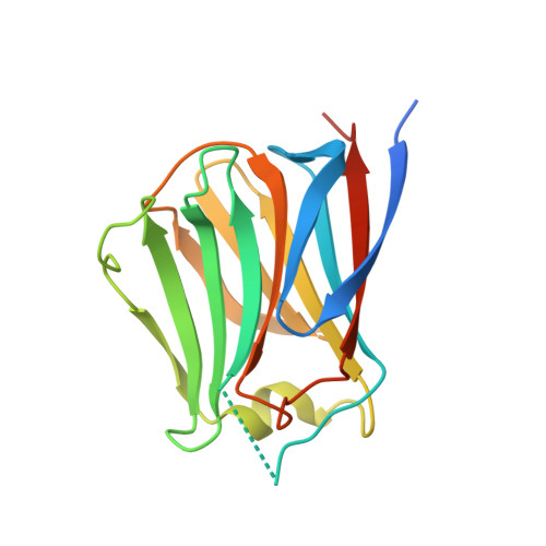
Conformational change of a unique sequence in a fungal galectin from Agrocybe cylindracea controls glycan ligand-binding specificity.

Kuwabara, N.Hu, D.Tateno, H.Makyio, H.Hirabayashi, J.Kato, R.

(2013) FEBS Lett 587: 3620-3625

  • DOI: https://doi.org/10.1016/j.febslet.2013.08.046
  • Primary Citation of Related Structures:  
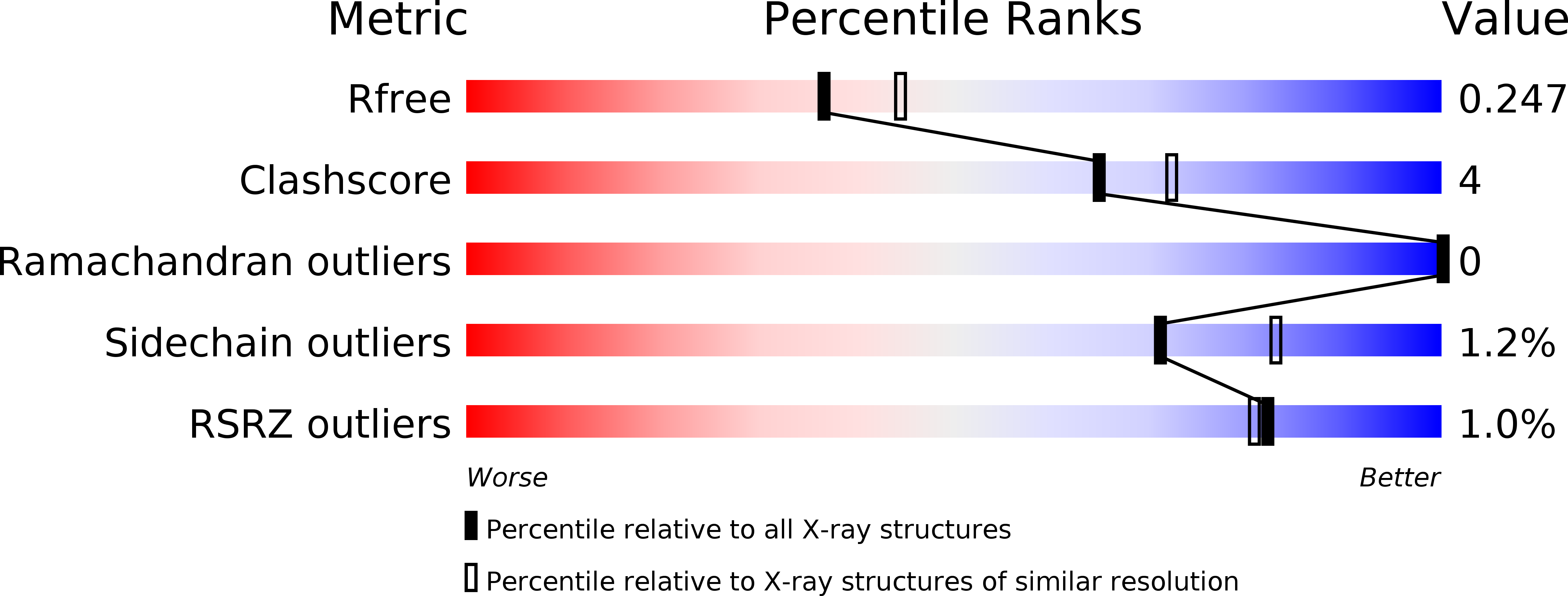
    3WG1, 3WG2, 3WG3, 3WG4

  • PubMed Abstract: 

    A fungal galectin from Agrocybe cylindracea (ACG) exhibits broad binding specificity for β-galactose-containing glycans. We determined the crystal structures of wild-type ACG and the N46A mutant, with and without glycan ligands. From these structures and a saccharide-binding analysis of the N46A mutant, we revealed that a conformational change of a unique insertion sequence containing Asn46 provides two binding modes for ACG, and thereby confers broad binding specificity. We propose that the unique sequence provides these two distinct glycan-binding modes by an induced-fit mechanism.


  • Organizational Affiliation

    Structural Biology Research Center, Photon Factory, Institute of Materials Structure Science, High Energy Accelerator Research Organization (KEK), 1-1 Oho, Tsukuba, Ibaraki 305-0801, Japan.


Macromolecules
Find similar proteins by:  (by identity cutoff)  |  3D Structure
Entity ID: 1
MoleculeChains Sequence LengthOrganismDetailsImage
Galactoside-binding lectin
A, B
178Cyclocybe aegeritaMutation(s): 0 
UniProt
Find proteins for Q6WY08 (Cyclocybe aegerita)
Explore Q6WY08 
Go to UniProtKB:  Q6WY08
Entity Groups  
Sequence Clusters30% Identity50% Identity70% Identity90% Identity95% Identity100% Identity
UniProt GroupQ6WY08
Sequence Annotations
Expand
  • Reference Sequence
Small Molecules
Ligands 1 Unique
IDChains Name / Formula / InChI Key2D Diagram3D Interactions
PGE
Query on PGE

Download Ideal Coordinates CCD File 
C [auth B]TRIETHYLENE GLYCOL
C6 H14 O4
ZIBGPFATKBEMQZ-UHFFFAOYSA-N
Experimental Data & Validation

Experimental Data

  • Method: X-RAY DIFFRACTION
  • Resolution: 2.20 Å
  • R-Value Free: 0.244 
  • R-Value Work: 0.211 
  • R-Value Observed: 0.213 
  • Space Group: P 61
Unit Cell:
Length ( Å )Angle ( ˚ )
a = 124.925α = 90
b = 124.925β = 90
c = 56.967γ = 120
Software Package:
Software NamePurpose
DENZOdata reduction
SCALEPACKdata scaling
REFMACrefinement
PDB_EXTRACTdata extraction
HKL-2000data collection
HKL-2000data reduction
HKL-2000data scaling
MOLREPphasing

Structure Validation

View Full Validation Report


Entry History 

Deposition Data

Revision History  (Full details and data files)

  • Version 1.0: 2013-11-27
    Type: Initial release
  • Version 1.1: 2017-11-22
    Changes: Advisory, Refinement description
  • Version 1.2: 2023-11-08
    Changes: Advisory, Data collection, Database references, Derived calculations, Refinement description