Role of a cysteine residue in the active site of ERK and the MAPKK family
Ohori, M., Kinoshita, T., Yoshimura, S., Warizaya, M., Nakajima, H., Miyake, H.(2007) Biochem Biophys Res Commun 353: 633-637
- PubMed: 17194451
- DOI: https://doi.org/10.1016/j.bbrc.2006.12.083
- Primary Citation of Related Structures:
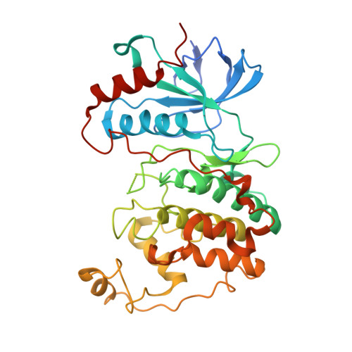
3W55 - PubMed Abstract:
Kinases of mitogen-activated protein kinase (MAPK) cascades, including extracellular signal-regulated protein kinase (ERK), represent likely targets for pharmacological intervention in proliferative diseases. Here, we report that FR148083 inhibits ERK2 enzyme activity and TGFbeta-induced AP-1-dependent luciferase expression with respective IC50 values of 0.08 and 0.05 microM. FR265083 (1'-2' dihydro form) and FR263574 (1'-2' and 7'-8' tetrahydro form) exhibited 5.5-fold less and no activity, respectively, indicating that both the alpha,beta-unsaturated ketone and the conformation of the lactone ring contribute to this inhibitory activity. The X-ray crystal structure of the ERK2/FR148083 complex revealed that the compound binds to the ATP binding site of ERK2, involving a covalent bond to Sgamma of ERK2 Cys166, hydrogen bonds with the backbone NH of Met108, Nzeta of Lys114, backbone C=O of Ser153, Ndelta2 of Asn154, and hydrophobic interactions with the side chains of Ile31, Val39, Ala52, and Leu156. The covalent bond motif in the ERK2/FR148083 complex assures that the inhibitor has high activity for ERK2 and no activity for other MAPKs such as JNK1 and p38MAPKalpha/beta/gamma/delta which have leucine residues at the site corresponding to Cys166 in ERK2. On the other hand, MEK1 and MKK7, kinases of the MAPKK family which also can be inhibited by FR148083, contain a cysteine residue corresponding to Cys166 of ERK2. The covalent binding to the common cysteine residue in the ATP-binding site is therefore likely to play a crucial role in the inhibitory activity for these MAP kinases. These findings on the molecular recognition mechanisms of FR148083 for kinases with Cys166 should provide a novel strategy for the pharmacological intervention of MAPK cascades.
Organizational Affiliation:
Astellas Pharma Inc., Tokodai 5-2-3, Tsukuba, Ibaraki 300-2698, Japan.