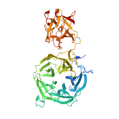
Crystal structure of 1,3Gal43A, an exo-beta-1,3-galactanase from Clostridium thermocellum
Jiang, D., Fan, J., Wang, X., Zhao, Y., Huang, B., Liu, J., Zhang, X.C.(2012) J Struct Biol 180: 447-457
- PubMed: 22960181
- DOI: https://doi.org/10.1016/j.jsb.2012.08.005
- Primary Citation of Related Structures:
3VSF, 3VSZ, 3VT0, 3VT1, 3VT2 - PubMed Abstract:
Glycoside hydrolase family 43 (GH43) consists of a variety of enzymes distributed widely in prokaryotes and eukaryotes. The mechanism by which GH43 enzymes hydrolyze oligosaccharides requires three essential acidic amino acid residues. However, one of them is thought to be missing in galactan β-1,3-galactosidases from the GH43 family. Ct1,3Gal43A, from Clostridium thermocellum, is comprised of a GH43 domain, a CBM13 domain, and a dockerin domain and exhibits an unusual ability to hydrolyze β-1,3-galactan in the presence of a β-1,6 linked branch. Here, we present its crystal structure at 2.7 Å resolution and complex structures of the enzyme with several substrates and analogs. Two modes of substrate binding were observed at the β site of the CtCBM13 domain, and one galactobiose molecule was found in an "L" shaped pocket of the CtGH43 domain, which appears large enough to accommodate two more galactose units. In addition, we found that mutating Glu112 to Gln or Ala eliminated the galactan hydrolysis activity of Ct1,3Gal43A while did not disrupt its ligand binding ability. Combining this results and the crystal structure we identified Glu112 in Ct1,3Gal43A as the 'missing' essential acidic residue in galactan β-1,3-galactosidases. Structural information presented here also suggests a mechanism by which Ct1,3Gal43A bypasses β-1,6 linked branches in the substrate and another mechanism by which the substrate is delivered 'in trans' from the CBM13 domain to the catalytic GH43 domain.
Organizational Affiliation:
Sino-France Laboratory for Drug Screening, Key Laboratory of Molecular Biophysics of Ministry of Education, School of Life Science and Technology, Huazhong University of Science and Technology, Wuhan 430074, Hubei, China.