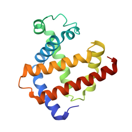
3VAU

Myoglobin nitrite structure: nitriheme modified


Experimental Data Snapshot

  • Method: X-RAY DIFFRACTION
  • Resolution: 1.70 Å
  • R-Value Free: 0.237 
  • R-Value Work: 0.173 
  • R-Value Observed: 0.176 

wwPDB Validation   3D Report Full Report


Ligand Structure Quality Assessment 


This is version 1.1 of the entry. See complete history


Literature

Unveiling the three-dimensional structure of the green pigment of nitrite-cured meat

Yi, J.Richter-Addo, G.B.

(2012) Chem Commun (Camb) 48: 4172-4174

  • DOI: https://doi.org/10.1039/c2cc31065a
  • Primary Citation of Related Structures:  
    3VAU

  • PubMed Abstract: 

    The 1.70 Å-resolution X-ray crystal structure of the green pigment occasionally present in nitrite-cured meat reveals a regiospecific nitration of the heme at the 2-vinyl position; the regiospecificity is favored on steric grounds, and results in movement of the 2-nitrovinyl moiety into the plane of the 24-atom heme macrocycle.


  • Organizational Affiliation

    Department of Chemistry and Biochemistry, University of Oklahoma, Norman, Oklahoma, USA. yijun@ou.edu


Macromolecules
Find similar proteins by:  (by identity cutoff)  |  3D Structure
Entity ID: 1
MoleculeChains Sequence LengthOrganismDetailsImage
Myoglobin153Equus caballusMutation(s): 0 
UniProt
Find proteins for P68082 (Equus caballus)
Explore P68082 
Go to UniProtKB:  P68082
Entity Groups  
Sequence Clusters30% Identity50% Identity70% Identity90% Identity95% Identity100% Identity
UniProt GroupP68082
Sequence Annotations
Expand
  • Reference Sequence
Small Molecules
Ligands 3 Unique
IDChains Name / Formula / InChI Key2D Diagram3D Interactions
NTE
Query on NTE

Download Ideal Coordinates CCD File 
B [auth A][3,3'-{7-ethenyl-3,8,13,17-tetramethyl-12-[(E)-2-nitroethenyl]porphyrin-2,18-diyl-kappa~4~N~21~,N~22~,N~23~,N~24~}dipro panoato(2-)]iron
C34 H31 Fe N5 O6
MBEUICWDKPMPCN-RXJYIKGQSA-L
SO4
Query on SO4

Download Ideal Coordinates CCD File 
D [auth A]SULFATE ION
O4 S
QAOWNCQODCNURD-UHFFFAOYSA-L
NO2
Query on NO2

Download Ideal Coordinates CCD File 
C [auth A]NITRITE ION
N O2
IOVCWXUNBOPUCH-UHFFFAOYSA-M
Experimental Data & Validation

Experimental Data

  • Method: X-RAY DIFFRACTION
  • Resolution: 1.70 Å
  • R-Value Free: 0.237 
  • R-Value Work: 0.173 
  • R-Value Observed: 0.176 
  • Space Group: P 1 21 1
Unit Cell:
Length ( Å )Angle ( ˚ )
a = 35.14α = 90
b = 28.423β = 105.5
c = 62.867γ = 90
Software Package:
Software NamePurpose
CrystalCleardata collection
PHASERphasing
REFMACrefinement
d*TREKdata reduction
d*TREKdata scaling

Structure Validation

View Full Validation Report


Ligand Structure Quality Assessment 


Entry History 

Deposition Data

Revision History  (Full details and data files)

  • Version 1.0: 2012-04-11
    Type: Initial release
  • Version 1.1: 2023-11-08
    Changes: Data collection, Database references, Derived calculations, Refinement description, Structure summary