3V21

Crystal structure of Type IIF restriction endonuclease Bse634I with cognate DNA


Experimental Data Snapshot

  • Method: X-RAY DIFFRACTION
  • Resolution: 2.70 Å
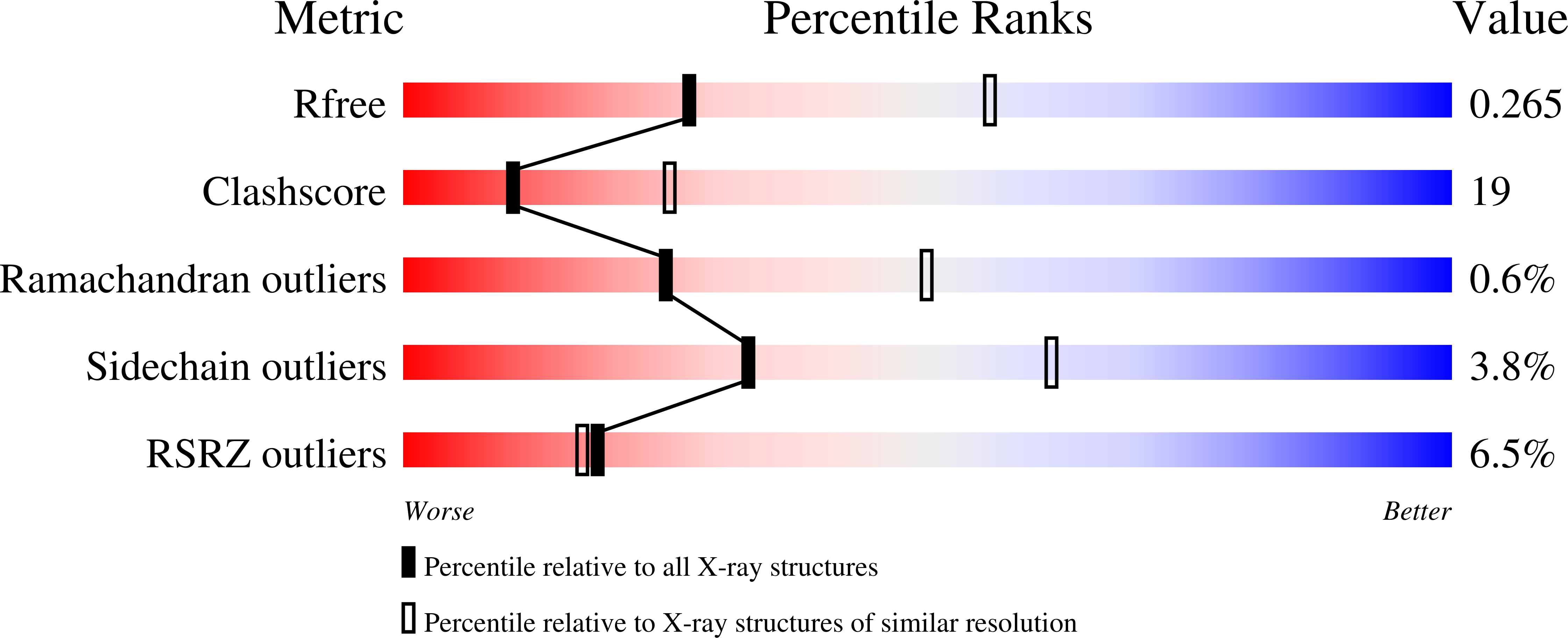
  • R-Value Free: 0.267 
  • R-Value Work: 0.230 
  • R-Value Observed: 0.234 

wwPDB Validation   3D Report Full Report


This is version 1.2 of the entry. See complete history


Literature

Structural mechanisms of the degenerate sequence recognition by Bse634I restriction endonuclease.

Manakova, E.Grazulis, S.Zaremba, M.Tamulaitiene, G.Golovenko, D.Siksnys, V.

(2012) Nucleic Acids Res 40: 6741-6751

  • DOI: https://doi.org/10.1093/nar/gks300
  • Primary Citation of Related Structures:  
    3V1Z, 3V20, 3V21

  • PubMed Abstract: 

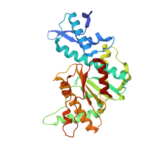
    Restriction endonuclease Bse634I recognizes and cleaves the degenerate DNA sequence 5'-R/CCGGY-3' (R stands for A or G; Y for T or C, '/' indicates a cleavage position). Here, we report the crystal structures of the Bse634I R226A mutant complexed with cognate oligoduplexes containing ACCGGT and GCCGGC sites, respectively. In the crystal, all potential H-bond donor and acceptor atoms on the base edges of the conserved CCGG core are engaged in the interactions with Bse634I amino acid residues located on the α6 helix. In contrast, direct contacts between the protein and outer base pairs are limited to van der Waals contact between the purine nucleobase and Pro203 residue in the major groove and a single H-bond between the O2 atom of the outer pyrimidine and the side chain of the Asn73 residue in the minor groove. Structural data coupled with biochemical experiments suggest that both van der Waals interactions and indirect readout contribute to the discrimination of the degenerate base pair by Bse634I. Structure comparison between related enzymes Bse634I (R/CCGGY), NgoMIV (G/CCGGC) and SgrAI (CR/CCGGYG) reveals how different specificities are achieved within a conserved structural core.


  • Organizational Affiliation

    Department of Protein-DNA Interactions, Institute of Biotechnology Vilnius University, Graiciuno 8, LT-02241 Vilnius, Lithuania.


Macromolecules

Find similar proteins by:  (by identity cutoff)  |  3D Structure
Entity ID: 1
MoleculeChains Sequence LengthOrganismDetailsImage
Endonuclease Bse634IR
A, B, C, D, E
A, B, C, D, E, F, G, H
293Geobacillus stearothermophilusMutation(s): 1 
Gene Names: bse634IR
EC: 3.1.21.4
UniProt
Find proteins for Q8RT53 (Geobacillus stearothermophilus)
Explore Q8RT53 
Go to UniProtKB:  Q8RT53
Entity Groups  
Sequence Clusters30% Identity50% Identity70% Identity90% Identity95% Identity100% Identity
UniProt GroupQ8RT53
Sequence Annotations
Expand
  • Reference Sequence

Find similar nucleic acids by:  Sequence   |   3D Structure  

Entity ID: 2
MoleculeChains LengthOrganismImage
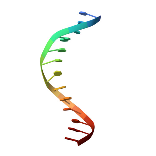
DNA (5'-D(*TP*TP*CP*GP*AP*CP*CP*GP*GP*TP*CP*GP*A)-3')13N/A
Sequence Annotations
Expand
  • Reference Sequence
Experimental Data & Validation

Experimental Data

  • Method: X-RAY DIFFRACTION
  • Resolution: 2.70 Å
  • R-Value Free: 0.267 
  • R-Value Work: 0.230 
  • R-Value Observed: 0.234 
  • Space Group: P 1 21 1
Unit Cell:
Length ( Å )Angle ( ˚ )
a = 106.88α = 90
b = 115.311β = 112.5
c = 130.243γ = 90
Software Package:
Software NamePurpose
REFMACrefinement
MOSFLMdata reduction
SCALAdata scaling
PDB_EXTRACTdata extraction
MAR345dtbdata collection
MOLREPphasing

Structure Validation

View Full Validation Report


Entry History 

Deposition Data

Revision History  (Full details and data files)

  • Version 1.0: 2012-04-25
    Type: Initial release
  • Version 1.1: 2012-11-14
    Changes: Database references
  • Version 1.2: 2023-09-13
    Changes: Data collection, Database references, Refinement description