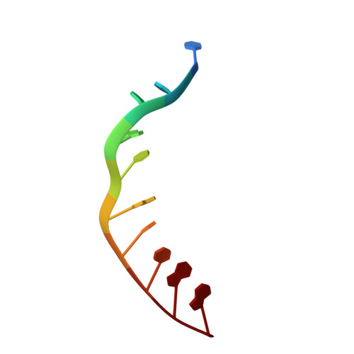
Crystal structure of lambda-[Ru(TAP)2(dppz)]2+ with d(TCGGTACCGA)2 displaying kinking of DNA via semiintercalation.
Niyazi, H.N., O'Sullivan, K., Texeira, S.C.M., Mitchell, E.P., Kelly, J.M., Cardin, J.C., Forsyth, V.T.To be published.
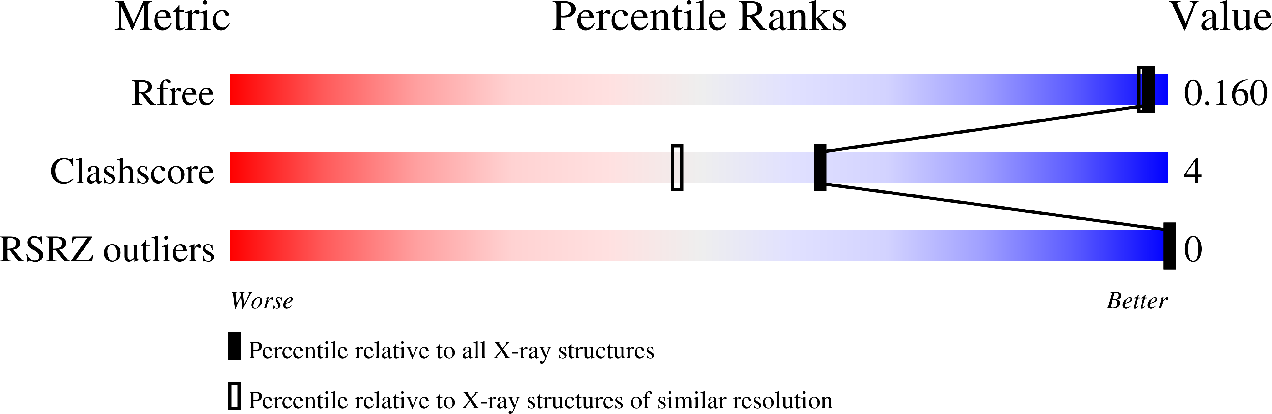
Experimental Data Snapshot
| Ligands 2 Unique | |||||
|---|---|---|---|---|---|
| ID | Chains | Name / Formula / InChI Key | 2D Diagram | 3D Interactions | |
| RKL Query on RKL | B [auth A], C [auth A] | Ru(tap)2(dppz) complex C38 H22 N12 Ru PIKVAZQLFXBUSD-UHFFFAOYSA-N |  | ||
| BA Query on BA | D [auth A] | BARIUM ION Ba XDFCIPNJCBUZJN-UHFFFAOYSA-N |  | ||
| Length ( Å ) | Angle ( ˚ ) |
|---|---|
| a = 54.376 | α = 90 |
| b = 54.376 | β = 90 |
| c = 28.995 | γ = 90 |
| Software Name | Purpose |
|---|---|
| MxCuBE | data collection |
| SHELXCD | phasing |
| SHELXE | model building |
| REFMAC | refinement |
| XDS | data reduction |
| SCALA | data scaling |
RCSB PDB (citation) is hosted by
RCSB PDB is a member of the