


Crystal Structure of Aba Receptor Pyl10
Sun, D.M., Wang, H.P., Wu, M.H., Zang, J.Y., Tian, C.L.To be published.
Experimental Data Snapshot
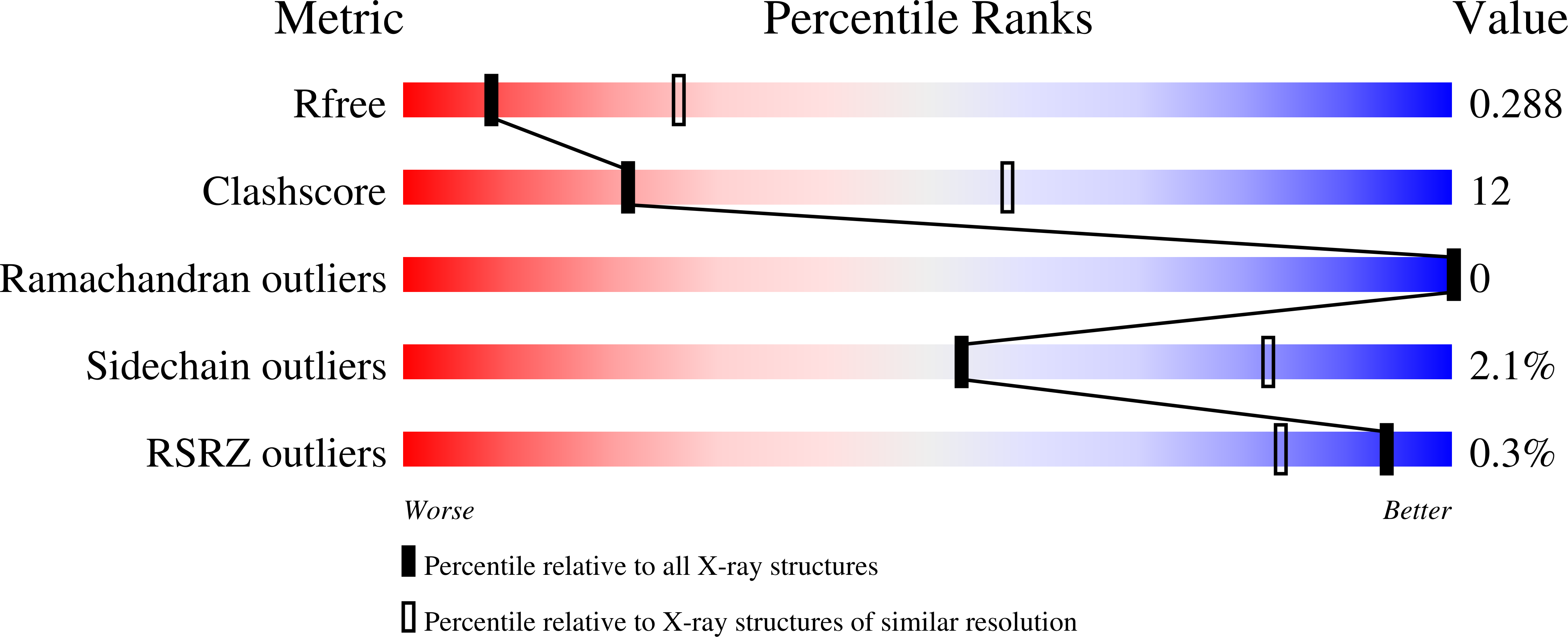
wwPDB Validation 3D Report Full Report
Entity ID: 1 | |||||
|---|---|---|---|---|---|
| Molecule | Chains | Sequence Length | Organism | Details | Image |
| Abscisic acid receptor PYL10 | 191 | Arabidopsis thaliana | Mutation(s): 0 Gene Names: AT4G27920, PYL10, RCAR4, T13J8.30 | 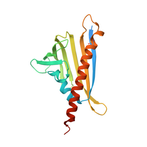 | |
UniProt | |||||
Find proteins for Q8H1R0 (Arabidopsis thaliana) Explore Q8H1R0 Go to UniProtKB: Q8H1R0 | |||||
Entity Groups | |||||
| Sequence Clusters | 30% Identity50% Identity70% Identity90% Identity95% Identity100% Identity | ||||
| UniProt Group | Q8H1R0 | ||||
Sequence AnnotationsExpand | |||||
| |||||
| Ligands 1 Unique | |||||
|---|---|---|---|---|---|
| ID | Chains | Name / Formula / InChI Key | 2D Diagram | 3D Interactions | |
| SO4 Query on SO4 | C [auth B] | SULFATE ION O4 S QAOWNCQODCNURD-UHFFFAOYSA-L |  | ||
| Length ( Å ) | Angle ( ˚ ) |
|---|---|
| a = 80.466 | α = 90 |
| b = 80.466 | β = 90 |
| c = 124.929 | γ = 120 |
| Software Name | Purpose |
|---|---|
| MAR345dtb | data collection |
| PHASER | phasing |
| REFMAC | refinement |
| HKL-2000 | data reduction |
| HKL-2000 | data scaling |
RCSB PDB (citation) is hosted by
RCSB PDB is a member of the

