3UJP

Structure of MntC protein at 2.7A


Experimental Data Snapshot

  • Method: X-RAY DIFFRACTION
  • Resolution: 2.70 Å
  • R-Value Free: 0.284 
  • R-Value Work: 0.231 
  • R-Value Observed: 0.233 

wwPDB Validation   3D Report Full Report


This is version 1.3 of the entry. See complete history


Literature

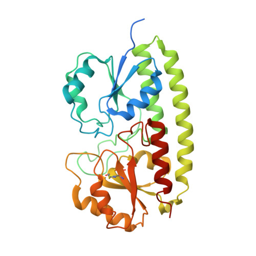
Arginine 116 stabilizes the entrance to the metal ion-binding site of the MntC protein.

Kanteev, M.Adir, N.

(2013) Acta Crystallogr Sect F Struct Biol Cryst Commun 69: 237-242

  • DOI: https://doi.org/10.1107/S174430911300153X
  • Primary Citation of Related Structures:  
    3UJP, 4IRM

  • PubMed Abstract: 

    The cyanobacterium Synechocystis sp. PCC 6803 imports Mn2+ ions via MntCAB, an ABC transport system that is expressed at submicromolar Mn2+ concentrations. The structures of the wild type (WT) and a site-directed mutant of the MntC solute-binding protein have been determined at 2.7 and 3.5 Å resolution, respectively. The WT structure is significantly improved over the previously determined structure (PDB entry 1xvl), showing improved Mn2+ binding site parameters, disulfide bonds in all three monomers and ions bound to the protein surface, revealing the role of Zn2+ ions in the crystallization liquor. The structure of MntC reveals that the active site is surrounded by neutral-to-positive electrostatic potential and is dominated by a network of polar interactions centred around Arg116. The mutation of this residue to alanine was shown to destabilize loops in the entrance to the metal-ion binding site and suggests a possible role in MntC function.


  • Organizational Affiliation

    Schulich Faculty of Chemistry, Technion-Israel Institute of Technology, Technion City, Haifa 32000, Israel.


Macromolecules
Find similar proteins by:  (by identity cutoff)  |  3D Structure
Entity ID: 1
MoleculeChains Sequence LengthOrganismDetailsImage
Mn transporter subunit
A, B, C
307Synechocystis sp. PCC 6803Mutation(s): 0 
Gene Names: mntCsll1598SYNGTS_1343
UniProt
Find proteins for Q79EF9 (Synechocystis sp. (strain PCC 6803 / Kazusa))
Explore Q79EF9 
Go to UniProtKB:  Q79EF9
Entity Groups  
Sequence Clusters30% Identity50% Identity70% Identity90% Identity95% Identity100% Identity
UniProt GroupQ79EF9
Sequence Annotations
Expand
  • Reference Sequence
Small Molecules
Ligands 3 Unique
IDChains Name / Formula / InChI Key2D Diagram3D Interactions
CAC
Query on CAC

Download Ideal Coordinates CCD File 
I [auth A],
J [auth A],
O [auth C],
P [auth C]
CACODYLATE ION
C2 H6 As O2
OGGXGZAMXPVRFZ-UHFFFAOYSA-M
ZN
Query on ZN

Download Ideal Coordinates CCD File 
E [auth A]
F [auth A]
G [auth A]
H [auth A]
M [auth C]
E [auth A],
F [auth A],
G [auth A],
H [auth A],
M [auth C],
N [auth C]
ZINC ION
Zn
PTFCDOFLOPIGGS-UHFFFAOYSA-N
MN
Query on MN

Download Ideal Coordinates CCD File 
D [auth A],
K [auth B],
L [auth C]
MANGANESE (II) ION
Mn
WAEMQWOKJMHJLA-UHFFFAOYSA-N
Experimental Data & Validation

Experimental Data

  • Method: X-RAY DIFFRACTION
  • Resolution: 2.70 Å
  • R-Value Free: 0.284 
  • R-Value Work: 0.231 
  • R-Value Observed: 0.233 
  • Space Group: P 31 2 1
Unit Cell:
Length ( Å )Angle ( ˚ )
a = 127.52α = 90
b = 127.52β = 90
c = 89.73γ = 120
Software Package:
Software NamePurpose
DNAdata collection
PHASERphasing
PHENIXrefinement
MOSFLMdata reduction
SCALAdata scaling

Structure Validation

View Full Validation Report


Entry History 

Deposition Data

Revision History  (Full details and data files)

  • Version 1.0: 2012-11-14
    Type: Initial release
  • Version 1.1: 2013-02-06
    Changes: Database references
  • Version 1.2: 2013-03-13
    Changes: Database references
  • Version 1.3: 2013-06-19
    Changes: Database references