3UBY

Crystal structure of human alklyadenine DNA glycosylase in a lower and higher-affinity complex with DNA


Experimental Data Snapshot

  • Method: X-RAY DIFFRACTION
  • Resolution: 2.00 Å
  • R-Value Free: 0.265 
  • R-Value Work: 0.219 
  • R-Value Observed: 0.222 

wwPDB Validation   3D Report Full Report


This is version 1.4 of the entry. See complete history


Literature

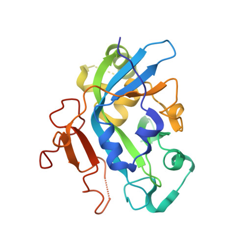
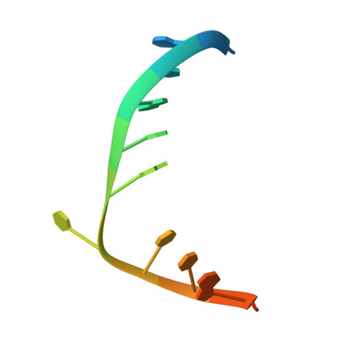
Searching for DNA lesions: structural evidence for lower- and higher-affinity DNA binding conformations of human alkyladenine DNA glycosylase.

Setser, J.W.Lingaraju, G.M.Davis, C.A.Samson, L.D.Drennan, C.L.

(2012) Biochemistry 51: 382-390

  • DOI: https://doi.org/10.1021/bi201484k
  • Primary Citation of Related Structures:  
    3UBY

  • PubMed Abstract: 

    To efficiently repair DNA, human alkyladenine DNA glycosylase (AAG) must search the million-fold excess of unmodified DNA bases to find a handful of DNA lesions. Such a search can be facilitated by the ability of glycosylases, like AAG, to interact with DNA using two affinities: a lower-affinity interaction in a searching process and a higher-affinity interaction for catalytic repair. Here, we present crystal structures of AAG trapped in two DNA-bound states. The lower-affinity depiction allows us to investigate, for the first time, the conformation of this protein in the absence of a tightly bound DNA adduct. We find that active site residues of AAG involved in binding lesion bases are in a disordered state. Furthermore, two loops that contribute significantly to the positive electrostatic surface of AAG are disordered. Additionally, a higher-affinity state of AAG captured here provides a fortuitous snapshot of how this enzyme interacts with a DNA adduct that resembles a one-base loop.


  • Organizational Affiliation

    Chemistry, Massachusetts Institute of Technology, Cambridge, Massachusetts 02139, United States.


Macromolecules

Find similar proteins by:  (by identity cutoff)  |  3D Structure
Entity ID: 1
MoleculeChains Sequence LengthOrganismDetailsImage
DNA-3-methyladenine glycosylase
A, B
219Homo sapiensMutation(s): 0 
Gene Names: MPGAAGANPGMID1
EC: 3.2.2.21
UniProt & NIH Common Fund Data Resources
Find proteins for P29372 (Homo sapiens)
Explore P29372 
Go to UniProtKB:  P29372
PHAROS:  P29372
GTEx:  ENSG00000103152 
Entity Groups  
Sequence Clusters30% Identity50% Identity70% Identity90% Identity95% Identity100% Identity
UniProt GroupP29372
Sequence Annotations
Expand
  • Reference Sequence

Find similar nucleic acids by:  Sequence   |   3D Structure  

Entity ID: 2
MoleculeChains LengthOrganismImage
DNA (5'-D(*GP*AP*CP*AP*TP*GP*(EDC)P*TP*TP*GP*CP*CP*T)-3')
C, D
13N/A
Sequence Annotations
Expand
  • Reference Sequence
Experimental Data & Validation

Experimental Data

  • Method: X-RAY DIFFRACTION
  • Resolution: 2.00 Å
  • R-Value Free: 0.265 
  • R-Value Work: 0.219 
  • R-Value Observed: 0.222 
  • Space Group: P 43
Unit Cell:
Length ( Å )Angle ( ˚ )
a = 41.168α = 90
b = 41.168β = 90
c = 262.546γ = 90
Software Package:
Software NamePurpose
ADSCdata collection
PHASERphasing
REFMACrefinement
DENZOdata reduction
SCALEPACKdata scaling

Structure Validation

View Full Validation Report


Entry History 

Deposition Data

Revision History  (Full details and data files)

  • Version 1.0: 2011-12-28
    Type: Initial release
  • Version 1.1: 2012-01-11
    Changes: Database references
  • Version 1.2: 2012-01-18
    Changes: Atomic model
  • Version 1.3: 2012-01-25
    Changes: Database references
  • Version 1.4: 2023-09-13
    Changes: Data collection, Database references, Derived calculations, Refinement description