3UBB

The crystal structure of GlpG in complex with a phosphonofluoridate inhibitor


Experimental Data Snapshot

  • Method: X-RAY DIFFRACTION
  • Resolution: 2.60 Å
  • R-Value Free: 0.229 
  • R-Value Work: 0.211 
  • R-Value Observed: 0.212 

wwPDB Validation   3D Report Full Report


Ligand Structure Quality Assessment 


This is version 1.1 of the entry. See complete history


Literature

Conformational Change in Rhomboid Protease GlpG Induced by Inhibitor Binding to Its S' Subsites.

Xue, Y.Chowdhury, S.Liu, X.Akiyama, Y.Ellman, J.Ha, Y.

(2012) Biochemistry 51: 3723-3731

  • DOI: https://doi.org/10.1021/bi300368b
  • Primary Citation of Related Structures:  
    3UBB

  • PubMed Abstract: 

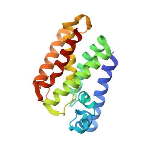
    Rhomboid protease conducts proteolysis inside the hydrophobic environment of the membrane. The conformational flexibility of the protease is essential for the enzyme mechanism, but the nature of this flexibility is not completely understood. Here we describe the crystal structure of rhomboid protease GlpG in complex with a phosphonofluoridate inhibitor, which is covalently bonded to the catalytic serine and extends into the S' side of the substrate binding cleft. Inhibitor binding causes subtle but extensive changes in the membrane protease. Many transmembrane helices tilt and shift positions, and the gap between S2 and S5 is slightly widened so that the inhibitor can bind between them. The side chain of Phe-245 from a loop (L5) that acts as a cap rotates and uncovers the opening of the substrate binding cleft to the lipid bilayer. A concurrent turn of the polypeptide backbone at Phe-245 moves the rest of the cap and exposes the catalytic serine to the aqueous solution. This study, together with earlier crystallographic investigation of smaller inhibitors, suggests a simple model for explaining substrate binding to rhomboid protease.


  • Organizational Affiliation

    Department of Pharmacology, Yale School of Medicine, New Haven, Connecticut 06520, USA.


Macromolecules
Find similar proteins by:  (by identity cutoff)  |  3D Structure
Entity ID: 1
MoleculeChains Sequence LengthOrganismDetailsImage
Rhomboid protease glpG182Escherichia coli K-12Mutation(s): 0 
Gene Names: glpGb3424JW5687
EC: 3.4.21.105
Membrane Entity: Yes 
UniProt
Find proteins for P09391 (Escherichia coli (strain K12))
Explore P09391 
Go to UniProtKB:  P09391
Entity Groups  
Sequence Clusters30% Identity50% Identity70% Identity90% Identity95% Identity100% Identity
UniProt GroupP09391
Sequence Annotations
Expand
  • Reference Sequence
Small Molecules
Ligands 1 Unique
IDChains Name / Formula / InChI Key2D Diagram3D Interactions
3UB
Query on 3UB

Download Ideal Coordinates CCD File 
B [auth A]propan-2-yl hydrogen (S)-[(1R)-1-{[(benzyloxy)carbonyl]amino}ethyl]phosphonate
C13 H20 N O5 P
DRLKMEFBZYZZJV-LLVKDONJSA-N
Experimental Data & Validation

Experimental Data

  • Method: X-RAY DIFFRACTION
  • Resolution: 2.60 Å
  • R-Value Free: 0.229 
  • R-Value Work: 0.211 
  • R-Value Observed: 0.212 
  • Space Group: H 3 2
Unit Cell:
Length ( Å )Angle ( ˚ )
a = 111.722α = 90
b = 111.722β = 90
c = 121.81γ = 120
Software Package:
Software NamePurpose
PHENIXrefinement
REFMACrefinement
CBASSdata collection
HKL-2000data reduction
HKL-2000data scaling
REFMACphasing

Structure Validation

View Full Validation Report


Ligand Structure Quality Assessment 


Entry History 

Deposition Data

  • Released Date: 2012-06-13 
  • Deposition Author(s): Xue, Y., Ha, Y.

Revision History  (Full details and data files)

  • Version 1.0: 2012-06-13
    Type: Initial release
  • Version 1.1: 2019-07-17
    Changes: Data collection, Derived calculations, Refinement description