3U30

Crystal structure of a linear-specific Ubiquitin fab bound to linear ubiquitin


Experimental Data Snapshot

  • Method: X-RAY DIFFRACTION
  • Resolution: 2.43 Å
  • R-Value Free: 0.258 
  • R-Value Work: 0.229 
  • R-Value Observed: 0.231 

wwPDB Validation   3D Report Full Report


This is version 1.3 of the entry. See complete history


Literature

Engineering and structural characterization of a linear polyubiquitin-specific antibody.

Matsumoto, M.L.Dong, K.C.Yu, C.Phu, L.Gao, X.Hannoush, R.N.Hymowitz, S.G.Kirkpatrick, D.S.Dixit, V.M.Kelley, R.F.

(2012) J Mol Biol 418: 134-144

  • DOI: https://doi.org/10.1016/j.jmb.2011.12.053
  • Primary Citation of Related Structures:  
    3U30

  • PubMed Abstract: 

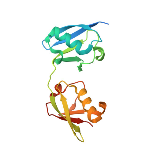
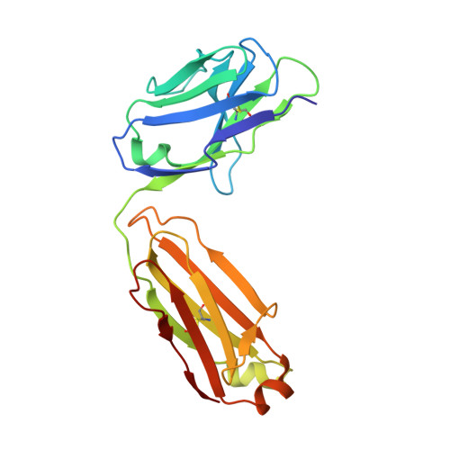
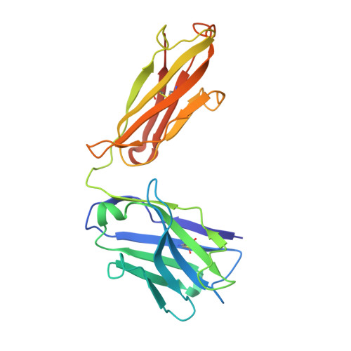
    Polyubiquitination is an essential posttranslational modification that plays critical roles in cellular signaling. PolyUb (polyubiquitin) chains are formed by linking the carboxyl-terminus of one Ub (ubiquitin) subunit to either a lysine residue or the amino-terminus of an adjacent Ub. Linkage through the amino-terminus results in linear polyubiquitination that has recently been demonstrated to be a key step in nuclear factor κB activation; however, tools to study linear chains have been lacking. We therefore engineered a linear-linkage-specific antibody that is functional in Western blot, immunoprecipitation, and immunofluorescence applications. A crystal structure of the linear-linkage-specific antibody Fab fragment in complex with linear diubiquitin provides molecular insight into the nature of linear chain specificity. We use the antibody to demonstrate that linear polyUb is up-regulated upon tumor necrosis factor α stimulation of cells, consistent with a critical role in nuclear factor κB signaling. This antibody provides an essential tool for further investigation of the function of linear chains.


  • Organizational Affiliation

    Department of Antibody Engineering, Genentech, Inc., 1 DNA Way, South San Francisco, CA 94080, USA.


Macromolecules
Find similar proteins by:  (by identity cutoff)  |  3D Structure
Entity ID: 1
MoleculeChains Sequence LengthOrganismDetailsImage
linear di-ubiquitin
A, D
172Homo sapiensMutation(s): 0 
Gene Names: UBC
UniProt & NIH Common Fund Data Resources
Find proteins for P0CG48 (Homo sapiens)
Explore P0CG48 
Go to UniProtKB:  P0CG48
PHAROS:  P0CG48
GTEx:  ENSG00000150991 
Entity Groups  
Sequence Clusters30% Identity50% Identity70% Identity90% Identity95% Identity100% Identity
UniProt GroupP0CG48
Sequence Annotations
Expand
  • Reference Sequence
Find similar proteins by:  (by identity cutoff)  |  3D Structure
Entity ID: 2
MoleculeChains Sequence LengthOrganismDetailsImage
Light chain Fab
B, E
214Homo sapiensMutation(s): 0 
Entity Groups  
Sequence Clusters30% Identity50% Identity70% Identity90% Identity95% Identity100% Identity
Sequence Annotations
Expand
  • Reference Sequence
Find similar proteins by:  (by identity cutoff)  |  3D Structure
Entity ID: 3
MoleculeChains Sequence LengthOrganismDetailsImage
Heavy chain Fab
C, F
227Homo sapiensMutation(s): 0 
Entity Groups  
Sequence Clusters30% Identity50% Identity70% Identity90% Identity95% Identity100% Identity
Sequence Annotations
Expand
  • Reference Sequence
Experimental Data & Validation

Experimental Data

  • Method: X-RAY DIFFRACTION
  • Resolution: 2.43 Å
  • R-Value Free: 0.258 
  • R-Value Work: 0.229 
  • R-Value Observed: 0.231 
  • Space Group: P 1
Unit Cell:
Length ( Å )Angle ( ˚ )
a = 53.322α = 87.08
b = 59.767β = 76.77
c = 96.033γ = 71.7
Software Package:
Software NamePurpose
BOSdata collection
PHASERphasing
PHENIXrefinement
HKL-2000data reduction
HKL-2000data scaling

Structure Validation

View Full Validation Report


Entry History 

Deposition Data

Revision History  (Full details and data files)

  • Version 1.0: 2012-01-18
    Type: Initial release
  • Version 1.1: 2013-01-09
    Changes: Database references
  • Version 1.2: 2018-01-24
    Changes: Structure summary
  • Version 1.3: 2023-09-13
    Changes: Data collection, Database references, Refinement description