3TOR

Crystal structure of Escherichia coli NrfA with Europium bound


Experimental Data Snapshot

  • Method: X-RAY DIFFRACTION
  • Resolution: 2.00 Å
  • R-Value Free: 0.222 
  • R-Value Work: 0.164 
  • R-Value Observed: 0.166 

wwPDB Validation   3D Report Full Report


Ligand Structure Quality Assessment 


This is version 1.1 of the entry. See complete history


Literature

Characterization of the active site and calcium binding in cytochrome c nitrite reductases.

Lockwood, C.W.Clarke, T.A.Butt, J.N.Hemmings, A.M.Richardson, D.J.

(2011) Biochem Soc Trans 39: 1871-1875

  • DOI: https://doi.org/10.1042/BST20110731
  • Primary Citation of Related Structures:  
    3TOR

  • PubMed Abstract: 

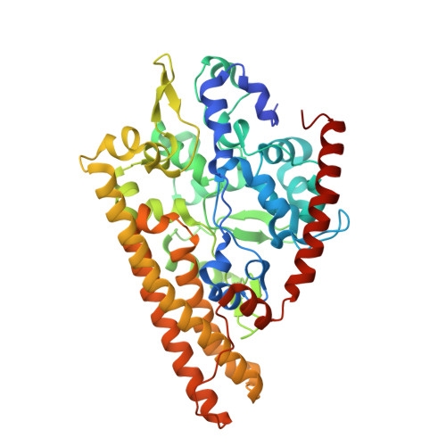
    The decahaem homodimeric cytochrome c nitrite reductase (NrfA) is expressed within the periplasm of a wide range of Gamma-, Delta- and Epsilon-proteobacteria and is responsible for the six-electron reduction of nitrite to ammonia. This allows nitrite to be used as a terminal electron acceptor, facilitating anaerobic respiration while allowing nitrogen to remain in a biologically available form. NrfA has also been reported to reduce nitric oxide (a reaction intermediate) and sulfite to ammonia and sulfide respectively, suggesting a potential secondary role as a detoxification enzyme. The protein sequences and crystal structures of NrfA from different bacteria and the closely related octahaem nitrite reductase from Thioalkalivibrio nitratireducens (TvNir) reveal that these enzymes are homologous. The NrfA proteins contain five covalently attached haem groups, four of which are bis-histidine-co-ordinated, with the proximal histidine being provided by the highly conserved CXXCH motif. These haems are responsible for intraprotein electron transfer. The remaining haem is the site for nitrite reduction, which is ligated by a novel lysine residue provided by a CXXCK haem-binding motif. The TvNir nitrite reductase has five haems that are structurally similar to those of NrfA and three extra bis-histidine-coordinated haems that precede the NrfA conserved region. The present review compares the protein sequences and structures of NrfA and TvNir and discusses the subtle differences related to active-site architecture and Ca2+ binding that may have an impact on substrate reduction.


  • Organizational Affiliation

    Centre for Molecular and Structural Biochemistry, School of Chemistry and School of Biological Sciences, University of East Anglia, Norwich Research Park, Norwich NR4 7TJ, UK.


Macromolecules
Find similar proteins by:  (by identity cutoff)  |  3D Structure
Entity ID: 1
MoleculeChains Sequence LengthOrganismDetailsImage
Cytochrome c nitrite reductase
A, B, C, D
452Escherichia coliMutation(s): 0 
EC: 1.7.2.2
UniProt
Find proteins for P0ABK9 (Escherichia coli (strain K12))
Explore P0ABK9 
Go to UniProtKB:  P0ABK9
Entity Groups  
Sequence Clusters30% Identity50% Identity70% Identity90% Identity95% Identity100% Identity
UniProt GroupP0ABK9
Sequence Annotations
Expand
  • Reference Sequence
Small Molecules
Ligands 3 Unique
IDChains Name / Formula / InChI Key2D Diagram3D Interactions
HEC
Query on HEC

Download Ideal Coordinates CCD File 
AA [auth C]
BA [auth C]
CA [auth C]
G [auth A]
GA [auth D]
AA [auth C],
BA [auth C],
CA [auth C],
G [auth A],
GA [auth D],
H [auth A],
HA [auth D],
I [auth A],
IA [auth D],
J [auth A],
JA [auth D],
K [auth A],
KA [auth D],
P [auth B],
Q [auth B],
R [auth B],
S [auth B],
T [auth B],
Y [auth C],
Z [auth C]
HEME C
C34 H34 Fe N4 O4
HXQIYSLZKNYNMH-LJNAALQVSA-N
EU
Query on EU

Download Ideal Coordinates CCD File 
DA [auth C]
F [auth A]
FA [auth D]
L [auth A]
M [auth A]
DA [auth C],
F [auth A],
FA [auth D],
L [auth A],
M [auth A],
O [auth B],
U [auth B],
V [auth B],
X [auth C]
EUROPIUM ION
Eu
MGVUQZZTJGLWJV-UHFFFAOYSA-N
CA
Query on CA

Download Ideal Coordinates CCD File 
E [auth A],
EA [auth D],
N [auth B],
W [auth C]
CALCIUM ION
Ca
BHPQYMZQTOCNFJ-UHFFFAOYSA-N
Experimental Data & Validation

Experimental Data

  • Method: X-RAY DIFFRACTION
  • Resolution: 2.00 Å
  • R-Value Free: 0.222 
  • R-Value Work: 0.164 
  • R-Value Observed: 0.166 
  • Space Group: P 21 21 21
Unit Cell:
Length ( Å )Angle ( ˚ )
a = 78.884α = 90
b = 90.489β = 90
c = 292.196γ = 90
Software Package:
Software NamePurpose
ADSCdata collection
MOLREPphasing
REFMACrefinement
DENZOdata reduction
SCALEPACKdata scaling

Structure Validation

View Full Validation Report


Ligand Structure Quality Assessment 


Entry History 

Deposition Data

Revision History  (Full details and data files)

  • Version 1.0: 2011-12-07
    Type: Initial release
  • Version 1.1: 2023-09-13
    Changes: Data collection, Database references, Derived calculations, Refinement description