3TO0

Crystal structure of Mus musculus iodotyrosine deiodinase (IYD) C217A, C239A bound to FMN


Experimental Data Snapshot

  • Method: X-RAY DIFFRACTION
  • Resolution: 2.65 Å
  • R-Value Free: 0.237 
  • R-Value Work: 0.182 
  • R-Value Observed: 0.185 

wwPDB Validation   3D Report Full Report


Ligand Structure Quality Assessment 


This is version 1.2 of the entry. See complete history


Literature

Expression of a soluble form of iodotyrosine deiodinase for active site characterization by engineering the native membrane protein from Mus musculus.

Buss, J.M.McTamney, P.M.Rokita, S.E.

(2012) Protein Sci 21: 351-361

  • DOI: https://doi.org/10.1002/pro.2020
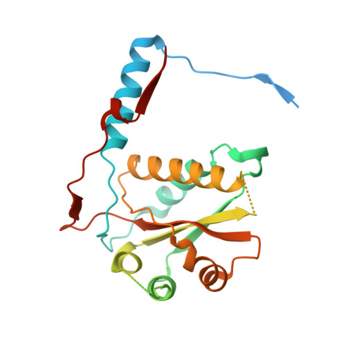
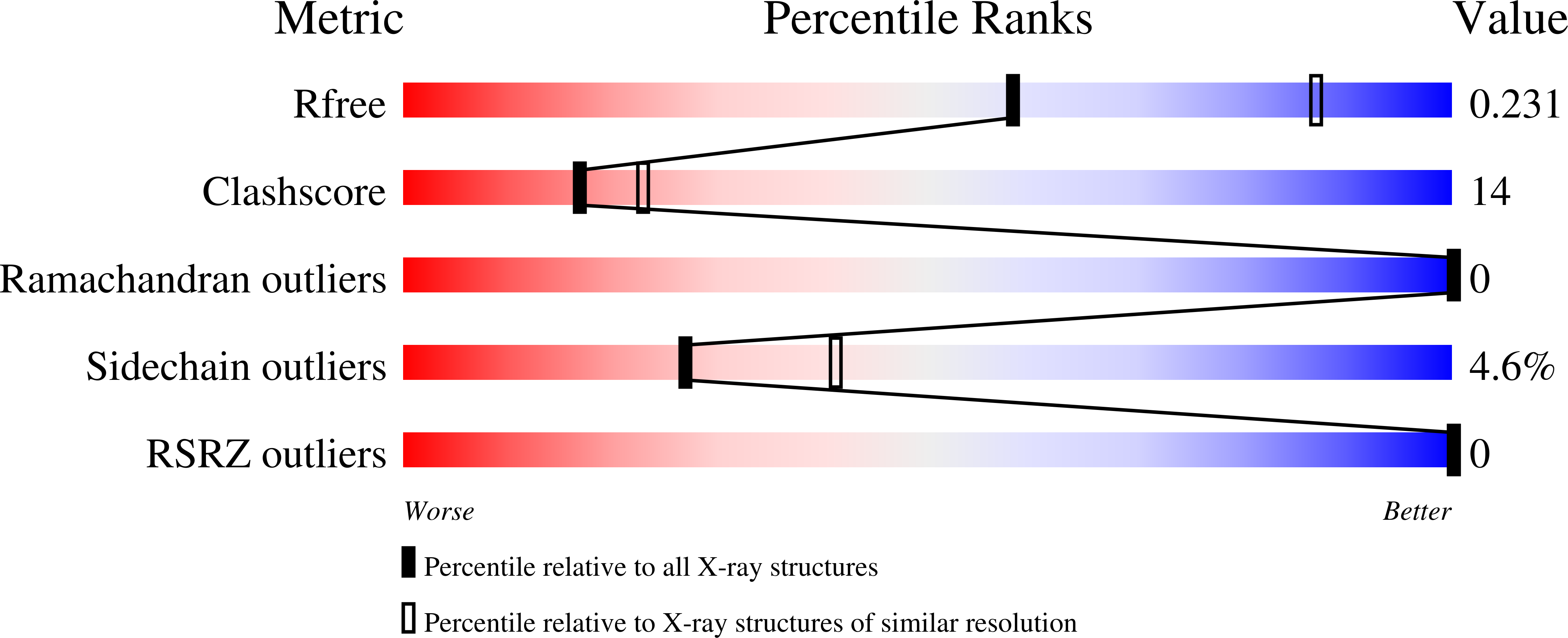
  • Primary Citation of Related Structures:  
    3TNZ, 3TO0

  • PubMed Abstract: 

    Reductive deiodination is critical for thyroid function and represents an unusual exception to the more common oxidative and hydrolytic mechanisms of dehalogenation in mammals. Studies on the reductive processes have been limited by a lack of convenient methods for heterologous expression of the appropriate proteins in large scale. The enzyme responsible for iodide salvage in the thyroid, iodotyrosine deodinase, is now readily generated after engineering its gene from Mus musculus. High expression of a truncated derivative lacking the membrane domain at its N-terminal was observed in Sf9 cells, whereas expression in Pichia pastoris remained low despite codon optimization. Ultimately, the desired expression in Escherichia coli was achieved after replacing the two conserved Cys residues of the deiodinase with Ala and fusing the resulting protein to thioredoxin. This final construct provided abundant enzyme for crystallography and mutagenesis. Utility of the E. coli system was demonstrated by examining a set of active site residues critical for binding to the zwitterionic portion of substrate.


  • Organizational Affiliation

    Department of Chemistry and Biochemistry, University of Maryland, College Park, Maryland 20742-2021, USA.


Macromolecules
Find similar proteins by:  (by identity cutoff)  |  3D Structure
Entity ID: 1
MoleculeChains Sequence LengthOrganismDetailsImage
Iodotyrosine deiodinase 1
A, B
259Mus musculusMutation(s): 2 
Gene Names: IYD-1
EC: 1.22.1.1
UniProt
Find proteins for Q9DCX8 (Mus musculus)
Explore Q9DCX8 
Go to UniProtKB:  Q9DCX8
Entity Groups  
Sequence Clusters30% Identity50% Identity70% Identity90% Identity95% Identity100% Identity
UniProt GroupQ9DCX8
Sequence Annotations
Expand
  • Reference Sequence
Experimental Data & Validation

Experimental Data

  • Method: X-RAY DIFFRACTION
  • Resolution: 2.65 Å
  • R-Value Free: 0.237 
  • R-Value Work: 0.182 
  • R-Value Observed: 0.185 
  • Space Group: P 31
Unit Cell:
Length ( Å )Angle ( ˚ )
a = 87.27α = 90
b = 87.27β = 90
c = 62.725γ = 120
Software Package:
Software NamePurpose
PROTEUM PLUSdata collection
CCP4model building
PHENIXrefinement
SAINTdata reduction
SADABSdata scaling
CCP4phasing

Structure Validation

View Full Validation Report


Ligand Structure Quality Assessment 


Entry History 

Deposition Data

Revision History  (Full details and data files)

  • Version 1.0: 2012-02-29
    Type: Initial release
  • Version 1.1: 2012-04-04
    Changes: Database references
  • Version 1.2: 2023-09-13
    Changes: Data collection, Database references, Derived calculations, Refinement description