3TMT

IrisFP, distorted chromophore


Experimental Data Snapshot

  • Method: X-RAY DIFFRACTION
  • Resolution: 2.00 Å
  • R-Value Free: 0.295 
  • R-Value Work: 0.295 
  • R-Value Observed: 0.295 

wwPDB Validation   3D Report Full Report


This is version 2.0 of the entry. See complete history


Literature

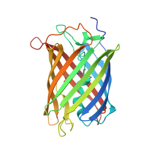
The nature of transient dark States in a photoactivatable fluorescent protein.

Roy, A.Field, M.J.Adam, V.Bourgeois, D.

(2011) J Am Chem Soc 133: 18586-18589

  • DOI: https://doi.org/10.1021/ja2085355
  • Primary Citation of Related Structures:  
    3TMR, 3TMT

  • PubMed Abstract: 

    Fluorescent proteins (FPs) of the green fluorescent protein family blink and bleach like all fluorophores. However, contrary to organic dyes, the mechanisms by which transient losses of fluorescence occur in FPs have received little attention. Here, we focus on the photoactivatable IrisFP, for which a transient non-fluorescent chromophoric state with distorted geometry was recently reported (Adam, V.; et al. J. Am. Chem. Soc. 009, 131, 18063). We investigated the chemical nature of this blinked state by employing quantum chemical/molecular mechanical calculations. Our findings suggest two previously unidentified dark states that display similar distorted chromophores with a transiently ruptured π-electron system. Both are protonated at atom C(α) of the chromophore methylene bridge. Transient protonation may occur via proton transfer from the nearby Arg66 either in the triplet state T(1) after intersystem crossing or in an anionic radical (doublet) ground state. As Arg66 is conserved in green-to-red photoconvertible FPs, these dark states are predicted to be common to all these proteins. We also suggest that C(α) protonated dark states may accelerate photobleaching by favoring decarboxylation of the fully conserved Glu212.


  • Organizational Affiliation

    Institut de Biologie Structurale (IBS)-Jean-Pierre Ebel, CEA/CNRS/ Université Joseph Fourier, 41, rue Jules Horowitz, 38027 Grenoble Cedex 1, France.


Macromolecules
Find similar proteins by:  (by identity cutoff)  |  3D Structure
Entity ID: 1
MoleculeChains Sequence LengthOrganismDetailsImage
Green to red photoconvertible GPF-like protein EosFP
A, B, C, D
230Lobophyllia hemprichiiMutation(s): 3 
UniProt
Find proteins for Q5S6Z9 (Lobophyllia hemprichii)
Explore Q5S6Z9 
Go to UniProtKB:  Q5S6Z9
Entity Groups  
Sequence Clusters30% Identity50% Identity70% Identity90% Identity95% Identity100% Identity
UniProt GroupQ5S6Z9
Sequence Annotations
Expand
  • Reference Sequence
Small Molecules
Ligands 1 Unique
IDChains Name / Formula / InChI Key2D Diagram3D Interactions
SO4
Query on SO4

Download Ideal Coordinates CCD File 
E [auth A]
F [auth A]
G [auth A]
H [auth A]
I [auth B]
E [auth A],
F [auth A],
G [auth A],
H [auth A],
I [auth B],
J [auth B],
K [auth B],
L [auth C],
M [auth C],
N [auth C],
O [auth C],
P [auth C],
Q [auth D],
R [auth D],
S [auth D],
T [auth D],
U [auth D]
SULFATE ION
O4 S
QAOWNCQODCNURD-UHFFFAOYSA-L
Modified Residues  1 Unique
IDChains TypeFormula2D DiagramParent
CR8
Query on CR8
A, B, C, D
L-PEPTIDE LINKINGC17 H16 N5 O4HIS, TYR, GLY
Experimental Data & Validation

Experimental Data

  • Method: X-RAY DIFFRACTION
  • Resolution: 2.00 Å
  • R-Value Free: 0.295 
  • R-Value Work: 0.295 
  • R-Value Observed: 0.295 
  • Space Group: P 21 21 21
Unit Cell:
Length ( Å )Angle ( ˚ )
a = 84.84α = 90
b = 96.51β = 90
c = 140γ = 90
Software Package:
Software NamePurpose
ADSCdata collection
PHASERphasing
PHENIXrefinement
XDSdata reduction
XSCALEdata scaling

Structure Validation

View Full Validation Report


Entry History 

Deposition Data

Revision History  (Full details and data files)

  • Version 1.0: 2011-11-16
    Type: Initial release
  • Version 1.1: 2011-11-23
    Changes: Structure summary
  • Version 1.2: 2011-11-30
    Changes: Database references
  • Version 2.0: 2023-11-15
    Changes: Advisory, Atomic model, Data collection, Database references, Derived calculations