3TC2

Crystal structure of potato serine protease inhibitor.


Experimental Data Snapshot

  • Method: X-RAY DIFFRACTION
  • Resolution: 1.60 Å
  • R-Value Free: 0.221 
  • R-Value Work: 0.168 
  • R-Value Observed: 0.171 

wwPDB Validation   3D Report Full Report


This is version 1.1 of the entry. See complete history


Literature

Structure of a post-translationally processed heterodimeric double-headed Kunitz-type serine protease inhibitor from potato.

Meulenbroek, E.M.Thomassen, E.A.Pouvreau, L.Abrahams, J.P.Gruppen, H.Pannu, N.S.

(2012) Acta Crystallogr D Biol Crystallogr 68: 794-799

  • DOI: https://doi.org/10.1107/S090744491201222X
  • Primary Citation of Related Structures:  
    3TC2

  • PubMed Abstract: 

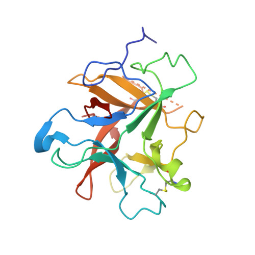
    Potato serine protease inhibitor (PSPI) constitutes about 22% of the total amount of proteins in potato tubers (cv. Elkana), making it the most abundant protease inhibitor in the plant. PSPI is a heterodimeric double-headed Kunitz-type serine protease inhibitor that can tightly and simultaneously bind two serine proteases by mimicking the substrate of the enzyme with its reactive-site loops. Here, the crystal structure of PSPI is reported, representing the first heterodimeric double-headed Kunitz-type serine protease inhibitor structure to be determined. PSPI has a β-trefoil fold and, based on the structure, two reactive-site loops bearing residues Phe75 and Lys95 were identified.


  • Organizational Affiliation

    Biophysical Structural Chemistry, Leiden University, Einsteinweg 55, 2333 CC Leiden, The Netherlands.


Macromolecules
Find similar proteins by:  (by identity cutoff)  |  3D Structure
Entity ID: 1
MoleculeChains Sequence LengthOrganismDetailsImage
Kunitz-type proteinase inhibitor P1H5
A, B, C
187Solanum tuberosumMutation(s): 0 
UniProt
Find proteins for Q8S380 (Solanum tuberosum)
Explore Q8S380 
Go to UniProtKB:  Q8S380
Entity Groups  
Sequence Clusters30% Identity50% Identity70% Identity90% Identity95% Identity100% Identity
UniProt GroupQ8S380
Sequence Annotations
Expand
  • Reference Sequence
Experimental Data & Validation

Experimental Data

  • Method: X-RAY DIFFRACTION
  • Resolution: 1.60 Å
  • R-Value Free: 0.221 
  • R-Value Work: 0.168 
  • R-Value Observed: 0.171 
  • Space Group: P 1 21 1
Unit Cell:
Length ( Å )Angle ( ˚ )
a = 54.822α = 90
b = 93.92β = 100.69
c = 55.442γ = 90
Software Package:
Software NamePurpose
ADSCdata collection
BALBESphasing
REFMACrefinement
MOSFLMdata reduction
SCALAdata scaling

Structure Validation

View Full Validation Report


Entry History 

Deposition Data

Revision History  (Full details and data files)

  • Version 1.0: 2012-06-20
    Type: Initial release
  • Version 1.1: 2014-04-16
    Changes: Database references