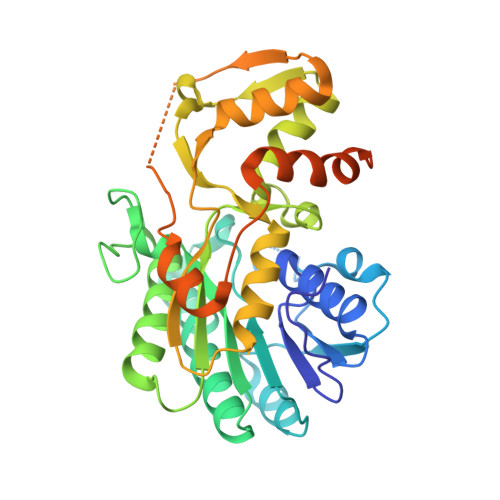
The crystal structure of ADP-L-glycero-D-manno-heptose-6-epimerase (HP0859) from Helicobacter pylori.
Shaik, M.M., Zanotti, G., Cendron, L.(2011) Biochim Biophys Acta 1814: 1641-1647
- PubMed: 21979583
- DOI: https://doi.org/10.1016/j.bbapap.2011.09.006
- Primary Citation of Related Structures:
3SXP - PubMed Abstract:
Helicobacter pylori, the human pathogen that affects about half of the world population and that is responsible for gastritis, gastric ulcer and adenocarcinoma and MALT lymphoma, owes much of the integrity of its outer membrane on lipopolysaccharides (LPSs). Together with their essential structural role, LPSs contribute to the bacterial adherence properties, as well as they are well characterized for the capability to modulate the immuno-response. In H. pylori the core oligosaccharide, one of the three main domains of LPSs, shows a peculiar structure in the branching organization of the repeating units, which displayed further variability when different strains have been compared. We present here the crystal structure of ADP-L-glycero-D-manno-heptose-6-epimerase (HP0859, rfaD), the last enzyme in the pathway that produces L-glycero-D-manno-heptose starting from sedoheptulose-7-phosphate, a crucial compound in the synthesis of the core oligosaccharide. In a recent study, a HP0859 knockout mutant has been characterized, demonstrating a severe loss of lipopolysaccharide structure and a significant reduction of adhesion levels in an infection model to AGS cells, if compared with the wild type strain, in good agreement with its enzymatic role. The crystal structure reveals that the enzyme is a homo-pentamer, and NAD is bound as a cofactor in a highly conserved pocket. The substrate-binding site of the enzyme is very similar to that of its orthologue in Escherichia coli, suggesting also a similar catalytic mechanism.
Organizational Affiliation:
Department of Biological Chemistry, University of Padua, Padua, Italy.