3SF4

Crystal structure of the complex between the conserved cell polarity proteins Inscuteable and LGN


Experimental Data Snapshot

  • Method: X-RAY DIFFRACTION
  • Resolution: 2.60 Å
  • R-Value Free: 0.262 
  • R-Value Work: 0.216 
  • R-Value Observed: 0.216 

wwPDB Validation   3D Report Full Report


This is version 1.2 of the entry. See complete history


Literature

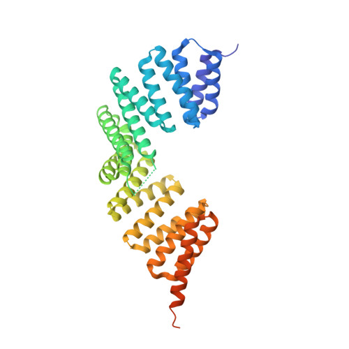
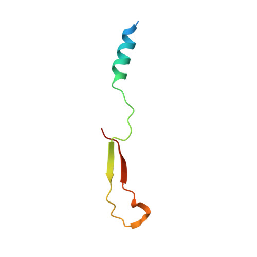
Structural basis for interaction between the conserved cell polarity proteins Inscuteable and Leu-Gly-Asn repeat-enriched protein (LGN)

Yuzawa, S.Kamakura, S.Iwakiri, Y.Hayase, J.Sumimoto, H.

(2011) Proc Natl Acad Sci U S A 108: 19210-19215

  • DOI: https://doi.org/10.1073/pnas.1110951108
  • Primary Citation of Related Structures:  
    3SF4

  • PubMed Abstract: 

    Interaction between the mammalian cell polarity proteins mInsc (mammalian homologue of Inscuteable) and Leu-Gly-Asn repeat-enriched protein (LGN), as well as that between their respective Drosophila homologues Inscuteable and Partner of Inscuteable (Pins), plays crucial roles in mitotic spindle orientation, a process contributing to asymmetric cell division. Here, we report a crystal structure of the LGN-binding domain (LBD) of human mInsc complexed with the N-terminal tetratricopeptide repeat (TPR) motifs of human LGN at 2.6-Å resolution. In the complex, mInsc-LBD adopts an elongated structure with three binding modules--an α-helix, an extended region, and a β-sheet connected with a loop--that runs antiparallel to LGN along the concave surface of the superhelix formed by the TPRs. Structural analysis and structure-based mutagenesis define residues that are critical for mInsc-LGN association, and reveal that the activator of G-protein signaling 3 (AGS3)-binding protein Frmpd1 [4.1/ezrin/radixin/moesin (FERM) and PSD-95/Dlg/ZO-1 (PDZ) domain-containing protein 1] and its relative Frmpd4 interact with LGN via a region homologous to a part of mInsc-LBD, whereas nuclear mitotic apparatus protein (NuMA) and the C terminus of LGN recognize the TPR domain in a manner different from that by mInsc. mInsc binds to LGN with the highest affinity (K(D) ≈ 2.4 nM) and effectively replaces the Frmpd proteins, NuMA, and the LGN C terminus, suggesting the priority of mInsc in binding to LGN. We also demonstrate, using mutant proteins, that mInsc-LGN interaction is vital for stabilization of LGN and for intracellular localization of mInsc.


  • Organizational Affiliation

    Department of Biochemistry, Kyushu University Graduate School of Medical Sciences, Fukuoka 812-8582, Japan.


Macromolecules
Find similar proteins by:  (by identity cutoff)  |  3D Structure
Entity ID: 1
MoleculeChains Sequence LengthOrganismDetailsImage
G-protein-signaling modulator 2
A, B, C
406Homo sapiensMutation(s): 0 
Gene Names: LGN
UniProt & NIH Common Fund Data Resources
Find proteins for P81274 (Homo sapiens)
Explore P81274 
Go to UniProtKB:  P81274
PHAROS:  P81274
GTEx:  ENSG00000121957 
Entity Groups  
Sequence Clusters30% Identity50% Identity70% Identity90% Identity95% Identity100% Identity
UniProt GroupP81274
Sequence Annotations
Expand
  • Reference Sequence
Find similar proteins by:  (by identity cutoff)  |  3D Structure
Entity ID: 2
MoleculeChains Sequence LengthOrganismDetailsImage
Protein inscuteable homolog
D, E, F
52Homo sapiensMutation(s): 0 
Gene Names: INSC
UniProt & NIH Common Fund Data Resources
Find proteins for Q1MX18 (Homo sapiens)
Explore Q1MX18 
Go to UniProtKB:  Q1MX18
PHAROS:  Q1MX18
GTEx:  ENSG00000188487 
Entity Groups  
Sequence Clusters30% Identity50% Identity70% Identity90% Identity95% Identity100% Identity
UniProt GroupQ1MX18
Sequence Annotations
Expand
  • Reference Sequence
Experimental Data & Validation

Experimental Data

  • Method: X-RAY DIFFRACTION
  • Resolution: 2.60 Å
  • R-Value Free: 0.262 
  • R-Value Work: 0.216 
  • R-Value Observed: 0.216 
  • Space Group: P 41 21 2
Unit Cell:
Length ( Å )Angle ( ˚ )
a = 124.815α = 90
b = 124.815β = 90
c = 233.851γ = 90
Software Package:
Software NamePurpose
MOLREPphasing
PHASERphasing
CNSrefinement
HKL-2000data reduction
HKL-2000data scaling

Structure Validation

View Full Validation Report


Entry History 

Deposition Data

Revision History  (Full details and data files)

  • Version 1.0: 2011-11-23
    Type: Initial release
  • Version 1.1: 2013-06-19
    Changes: Database references
  • Version 1.2: 2023-11-01
    Changes: Data collection, Database references, Refinement description