3S18

Crystal structure of a plant albumin from cicer arietinum showing hemagglutination


Experimental Data Snapshot

  • Method: X-RAY DIFFRACTION
  • Resolution: 2.20 Å
  • R-Value Free: 0.223 
  • R-Value Work: 0.177 
  • R-Value Observed: 0.179 

wwPDB Validation   3D Report Full Report


This is version 1.2 of the entry. See complete history


Literature

Crystal structure of a plant albumin from Cicer arietinum (chickpea) possessing hemopexin fold and hemagglutination activity

Sharma, U.Katre, U.V.Suresh, C.G.

(2015) Planta 

  • DOI: https://doi.org/10.1007/s00425-014-2236-6
  • Primary Citation of Related Structures:  
    3S18, 3V6N

  • PubMed Abstract: 

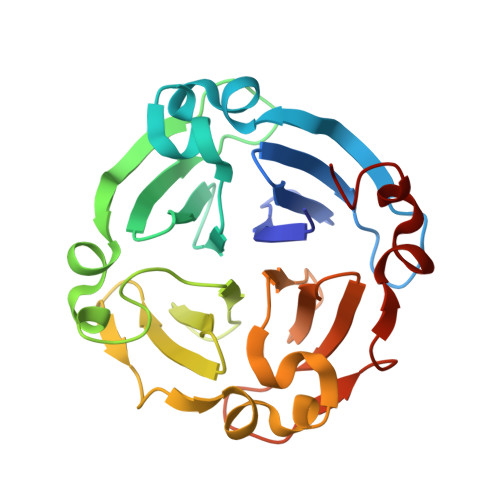
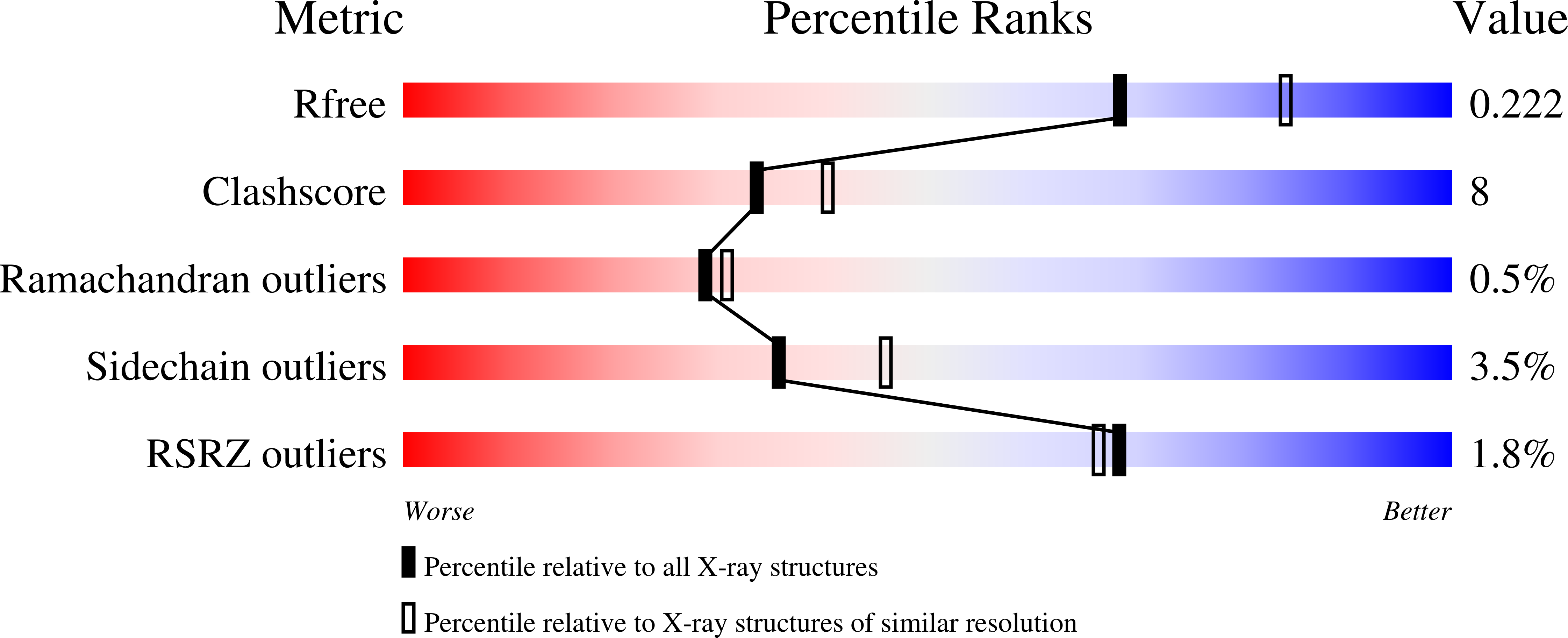
    Crystal structure of a reported PA2 albumin from Cicer arietinum shows that it belongs to hemopexin fold family, has four beta-propeller motifs and possesses hemagglutination activity, making it different from known legume lectins. A plant albumin (PA2) from Cicer arietinum, presumably a lectin (CAL) owing to its hemagglutination activity which is inhibited by complex sugars as well as glycoproteins such as fetuin, desialylated fetuin and fibrinogen. The three-dimensional structure of this homodimeric protein has been determined using X-ray crystallography at 2.2 Å in two crystal forms: orthorhombic (P21212) and trigonal (P3). The structure determined using molecular replacement method and refined in orthorhombic crystal form reached R-factors R free 22.6 % and R work 18.2 % and in trigonal form had 22.3 and 17.9 % in the resolution range of 20.0-2.2 and 35.3-2.2 Å, respectively. Interestingly, unlike the known legume lectin fold, the structure of this homodimeric hemagglutinin belonged to hemopexin fold that consisted of four-bladed β-propeller architecture. Each subunit has a central cavity forming a channel, inside of which is lined with hydrophobic residues. The channel also bears binding sites for ligands such as calcium, sodium and chloride ions, iodine atom in the case of iodine derivative and water molecules. However, none of these ligands seem important for the sugar recognition. No monosaccharide sugar specificity could be detected using hemagglutination inhibition. Chemical modification studies identified a potential sugar-binding site per subunit molecule. Comparison of C-alpha atom positions in subunit structures showed that the deviations between the two crystal forms were more with respect to blades I and IV. Differences also existed between subunits in two forms in terms of type and site of ligand binding.


  • Organizational Affiliation

    Division of Biochemical Sciences, CSIR-National Chemical Laboratory, Pune, 411008, India.


Macromolecules
Find similar proteins by:  (by identity cutoff)  |  3D Structure
Entity ID: 1
MoleculeChains Sequence LengthOrganismDetailsImage
lectin
A, B
227Cicer arietinumMutation(s): 0 
UniProt
Find proteins for G1K3S0 (Cicer arietinum)
Explore G1K3S0 
Go to UniProtKB:  G1K3S0
Entity Groups  
Sequence Clusters30% Identity50% Identity70% Identity90% Identity95% Identity100% Identity
UniProt GroupG1K3S0
Sequence Annotations
Expand
  • Reference Sequence
Small Molecules
Ligands 5 Unique
IDChains Name / Formula / InChI Key2D Diagram3D Interactions
IOD
Query on IOD

Download Ideal Coordinates CCD File 
G [auth A],
L [auth B]
IODIDE ION
I
XMBWDFGMSWQBCA-UHFFFAOYSA-M
SO4
Query on SO4

Download Ideal Coordinates CCD File 
C [auth A],
I [auth B]
SULFATE ION
O4 S
QAOWNCQODCNURD-UHFFFAOYSA-L
CA
Query on CA

Download Ideal Coordinates CCD File 
D [auth A],
J [auth B]
CALCIUM ION
Ca
BHPQYMZQTOCNFJ-UHFFFAOYSA-N
CL
Query on CL

Download Ideal Coordinates CCD File 
E [auth A],
F [auth A],
K [auth B]
CHLORIDE ION
Cl
VEXZGXHMUGYJMC-UHFFFAOYSA-M
NA
Query on NA

Download Ideal Coordinates CCD File 
H [auth A]SODIUM ION
Na
FKNQFGJONOIPTF-UHFFFAOYSA-N
Experimental Data & Validation

Experimental Data

  • Method: X-RAY DIFFRACTION
  • Resolution: 2.20 Å
  • R-Value Free: 0.223 
  • R-Value Work: 0.177 
  • R-Value Observed: 0.179 
  • Space Group: P 3
Unit Cell:
Length ( Å )Angle ( ˚ )
a = 81.917α = 90
b = 81.917β = 90
c = 69.591γ = 120
Software Package:
Software NamePurpose
CrystalCleardata collection
PHASERphasing
REFMACrefinement
DENZOdata reduction
SCALEPACKdata scaling

Structure Validation

View Full Validation Report


Entry History 

Deposition Data

Revision History  (Full details and data files)

  • Version 1.0: 2011-07-13
    Type: Initial release
  • Version 1.1: 2015-02-25
    Changes: Database references
  • Version 1.2: 2023-11-01
    Changes: Data collection, Database references, Derived calculations, Refinement description