3QYW

Crystal structure of ERK2 in complex with an inhibitor


Experimental Data Snapshot

  • Method: X-RAY DIFFRACTION
  • Resolution: 1.50 Å
  • R-Value Free: 0.188 
  • R-Value Work: 0.148 
  • R-Value Observed: 0.150 

wwPDB Validation   3D Report Full Report


Ligand Structure Quality Assessment 


This is version 1.2 of the entry. See complete history


Literature

In-plate protein crystallization, in situ ligand soaking and X-ray diffraction.

le Maire, A.Gelin, M.Pochet, S.Hoh, F.Pirocchi, M.Guichou, J.F.Ferrer, J.L.Labesse, G.

(2011) Acta Crystallogr D Biol Crystallogr 67: 747-755

  • DOI: https://doi.org/10.1107/S0907444911023249
  • Primary Citation of Related Structures:  
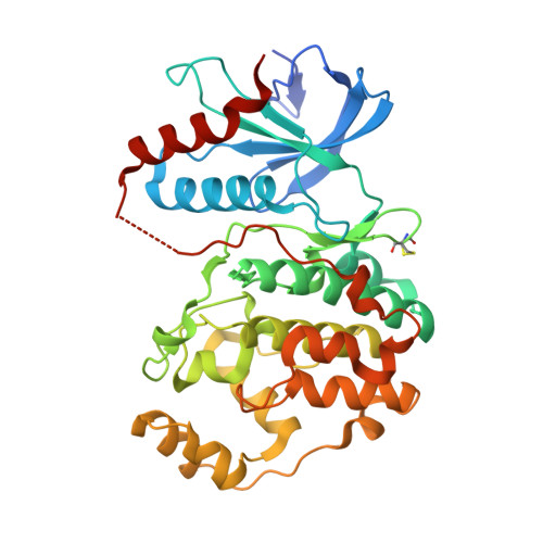
    3QYU, 3QYW, 3QYZ

  • PubMed Abstract: 

    X-ray crystallography is now a recognized technique for ligand screening, especially for fragment-based drug design. However, protein crystal handling is still tedious and limits further automation. An alternative method for the solution of crystal structures of proteins in complex with small ligands is proposed. Crystallization drops are directly exposed to an X-ray beam after cocrystallization or soaking with the desired ligands. The use of dedicated plates in connection with an optimal parametrization of the G-rob robot allows efficient data collection. Three proteins currently under study in our laboratory for ligand screening by X-ray crystallography were used as validation test cases. The protein crystals belonged to different space groups, including a challenging monoclinic case. The resulting diffraction data can lead to clear ligand recognition, including indication of alternating conformations. These results demonstrate a possible method for automation of ligand screening by X-ray crystallography.


  • Organizational Affiliation

    Université Montpellier 1 et 2, Centre de Biochimie Structurale, France.


Macromolecules
Find similar proteins by:  (by identity cutoff)  |  3D Structure
Entity ID: 1
MoleculeChains Sequence LengthOrganismDetailsImage
Mitogen-activated protein kinase 1364Rattus norvegicusMutation(s): 0 
Gene Names: Mapk1Erk2MapkPrkm1
EC: 2.7.11.24
UniProt
Find proteins for P63086 (Rattus norvegicus)
Explore P63086 
Go to UniProtKB:  P63086
Entity Groups  
Sequence Clusters30% Identity50% Identity70% Identity90% Identity95% Identity100% Identity
UniProt GroupP63086
Sequence Annotations
Expand
  • Reference Sequence
Small Molecules
Ligands 3 Unique
IDChains Name / Formula / InChI Key2D Diagram3D Interactions
6PB
Query on 6PB

Download Ideal Coordinates CCD File 
B [auth A]6-(3-bromophenyl)-7H-purin-2-amine
C11 H8 Br N5
OBKRZWFDMHKSFJ-UHFFFAOYSA-N
SO4
Query on SO4

Download Ideal Coordinates CCD File 
M [auth A]SULFATE ION
O4 S
QAOWNCQODCNURD-UHFFFAOYSA-L
DMS
Query on DMS

Download Ideal Coordinates CCD File 
C [auth A]
D [auth A]
E [auth A]
F [auth A]
G [auth A]
C [auth A],
D [auth A],
E [auth A],
F [auth A],
G [auth A],
H [auth A],
I [auth A],
J [auth A],
K [auth A],
L [auth A]
DIMETHYL SULFOXIDE
C2 H6 O S
IAZDPXIOMUYVGZ-UHFFFAOYSA-N
Modified Residues  1 Unique
IDChains TypeFormula2D DiagramParent
CME
Query on CME
A
L-PEPTIDE LINKINGC5 H11 N O3 S2CYS
Experimental Data & Validation

Experimental Data

  • Method: X-RAY DIFFRACTION
  • Resolution: 1.50 Å
  • R-Value Free: 0.188 
  • R-Value Work: 0.148 
  • R-Value Observed: 0.150 
  • Space Group: P 1 21 1
Unit Cell:
Length ( Å )Angle ( ˚ )
a = 49.067α = 90
b = 70.254β = 108.72
c = 60.168γ = 90
Software Package:
Software NamePurpose
ADSCdata collection
PHENIXrefinement
MOSFLMdata reduction
SCALAdata scaling

Structure Validation

View Full Validation Report


Ligand Structure Quality Assessment 


Entry History 

Deposition Data

Revision History  (Full details and data files)

  • Version 1.0: 2011-08-24
    Type: Initial release
  • Version 1.1: 2012-03-21
    Changes: Database references
  • Version 1.2: 2017-11-08
    Changes: Advisory, Refinement description