Cooperative macromolecular device revealed by meta-analysis of static and time-resolved structures.
Ren, Z., Srajer, V., Knapp, J.E., Royer Jr., W.E.(2012) Proc Natl Acad Sci U S A 109: 107-112
- PubMed: 22171006
- DOI: https://doi.org/10.1073/pnas.1109213108
- Primary Citation of Related Structures:
3QOB, 3UGY, 3UGZ, 3UH3, 3UH5, 3UH6, 3UH7, 3UHB, 3UHC, 3UHD, 3UHE, 3UHG, 3UHH, 3UHI, 3UHK, 3UHN, 3UHQ, 3UHR, 3UHS, 3UHT, 3UHU, 3UHV, 3UHW, 3UHX, 3UHY, 3UHZ, 3UI0 - PubMed Abstract:
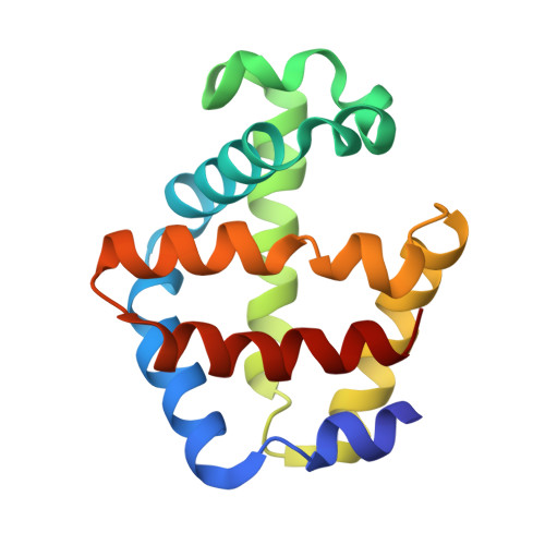
Here we present a meta-analysis of a large collection of static structures of a protein in the Protein Data Bank in order to extract the progression of structural events during protein function. We apply this strategy to the homodimeric hemoglobin HbI from Scapharca inaequivalvis. We derive a simple dynamic model describing how binding of the first ligand in one of the two chemically identical subunits facilitates a second binding event in the other partner subunit. The results of our ultrafast time-resolved crystallographic studies support this model. We demonstrate that HbI functions like a homodimeric mechanical device, such as pliers or scissors. Ligand-induced motion originating in one subunit is transmitted to the other via conserved pivot points, where the E and F' helices from two partner subunits are "bolted" together to form a stable dimer interface permitting slight relative rotation but preventing sliding.
Organizational Affiliation:
Center for Advanced Radiation Sources, University of Chicago, 9700 South Cass Avenue, Building 434B, Argonne, IL 60439, USA. renz@uchicago.edu