3QKT

Rad50 ABC-ATPase with adjacent coiled-coil region in complex with AMP-PNP


Experimental Data Snapshot

  • Method: X-RAY DIFFRACTION
  • Resolution: 1.90 Å
  • R-Value Free: 0.244 
  • R-Value Work: 0.192 
  • R-Value Observed: 0.195 

wwPDB Validation   3D Report Full Report


Ligand Structure Quality Assessment 


This is version 1.3 of the entry. See complete history


Literature

ABC ATPase signature helices in Rad50 link nucleotide state to Mre11 interface for DNA repair.

Williams, G.J.Williams, R.S.Williams, J.S.Moncalian, G.Arvai, A.S.Limbo, O.Guenther, G.Sildas, S.Hammel, M.Russell, P.Tainer, J.A.

(2011) Nat Struct Mol Biol 18: 423-431

  • DOI: https://doi.org/10.1038/nsmb.2038
  • Primary Citation of Related Structures:  
    3QKR, 3QKS, 3QKT, 3QKU

  • PubMed Abstract: 

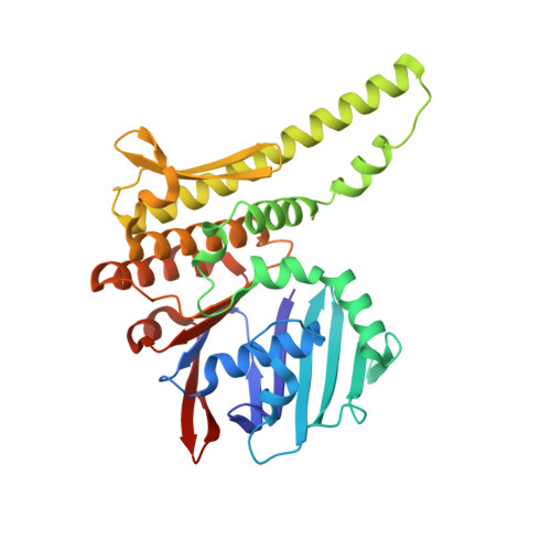
    The Rad50 ABC-ATPase complex with Mre11 nuclease is essential for dsDNA break repair, telomere maintenance and ataxia telangiectasia-mutated kinase checkpoint signaling. How Rad50 affects Mre11 functions and how ABC-ATPases communicate nucleotide binding and ligand states across long distances and among protein partners are questions that have remained obscure. Here, structures of Mre11-Rad50 complexes define the Mre11 2-helix Rad50 binding domain (RBD) that forms a four-helix interface with Rad50 coiled coils adjoining the ATPase core. Newly identified effector and basic-switch helix motifs extend the ABC-ATPase signature motif to link ATP-driven Rad50 movements to coiled coils binding Mre11, implying an ~30-Å pull on the linker to the nuclease domain. Both RBD and basic-switch mutations cause clastogen sensitivity. Our new results characterize flexible ATP-dependent Mre11 regulation, defects in cancer-linked RBD mutations, conserved superfamily basic switches and motifs effecting ATP-driven conformational change, and they provide a unified comprehension of ABC-ATPase activities.


  • Organizational Affiliation

    Life Sciences Division, Lawrence Berkeley National Laboratory, Berkeley, California, USA.


Macromolecules
Find similar proteins by:  (by identity cutoff)  |  3D Structure
Entity ID: 1
MoleculeChains Sequence LengthOrganismDetailsImage
DNA double-strand break repair rad50 ATPase
A, B, C, D
339Pyrococcus furiosusMutation(s): 0 
Gene Names: rad50PF1167
EC: 3.6
UniProt
Find proteins for P58301 (Pyrococcus furiosus (strain ATCC 43587 / DSM 3638 / JCM 8422 / Vc1))
Explore P58301 
Go to UniProtKB:  P58301
Entity Groups  
Sequence Clusters30% Identity50% Identity70% Identity90% Identity95% Identity100% Identity
UniProt GroupP58301
Sequence Annotations
Expand
  • Reference Sequence
Experimental Data & Validation

Experimental Data

  • Method: X-RAY DIFFRACTION
  • Resolution: 1.90 Å
  • R-Value Free: 0.244 
  • R-Value Work: 0.192 
  • R-Value Observed: 0.195 
  • Space Group: P 21 21 21
Unit Cell:
Length ( Å )Angle ( ˚ )
a = 83.39α = 90
b = 108.517β = 90
c = 150.366γ = 90
Software Package:
Software NamePurpose
Blu-Icedata collection
PHASERphasing
PHENIXrefinement
HKL-2000data reduction
HKL-2000data scaling

Structure Validation

View Full Validation Report


Ligand Structure Quality Assessment 


Entry History 

Deposition Data

Revision History  (Full details and data files)

  • Version 1.0: 2011-03-30
    Type: Initial release
  • Version 1.1: 2011-07-13
    Changes: Version format compliance
  • Version 1.2: 2017-07-26
    Changes: Refinement description, Source and taxonomy
  • Version 1.3: 2023-09-13
    Changes: Data collection, Database references, Derived calculations, Refinement description