3QJM

Structural flexibility of Shank PDZ domain is important for its binding to different ligands


Experimental Data Snapshot

  • Method: X-RAY DIFFRACTION
  • Resolution: 2.31 Å
  • R-Value Free: 0.267 
  • R-Value Work: 0.235 
  • R-Value Observed: 0.236 

wwPDB Validation   3D Report Full Report


This is version 1.1 of the entry. See complete history


Literature

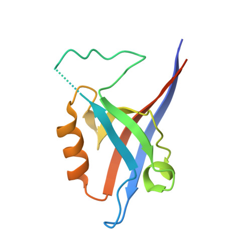
The structural flexibility of the shank1 PDZ domain is important for its binding to different ligands

Lee, J.H.Park, H.Park, S.J.Kim, H.J.Eom, S.H.

(2011) Biochem Biophys Res Commun 407: 207-212

  • DOI: https://doi.org/10.1016/j.bbrc.2011.02.141
  • Primary Citation of Related Structures:  
    3QJM, 3QJN

  • PubMed Abstract: 

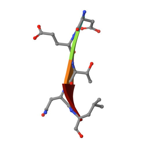
    The PDZ domain of the shank protein interacts with numerous cell membrane receptors and cytosolic proteins via the loosely defined binding motif X-(Ser/Thr)-X-Φ-COOH (Φ represents hydrophobic residues) at the carboxyl terminus of its target protein. This enables shank to serve as a membrane-associated scaffold for the assembly of signaling complexes. As the list of proteins that bind to the shank PDZ domain grows, it is not immediately clear what structural element(s) mediate this domain's target specificity or the plasticity required to bind its different targets. Here, we have determined the crystal structure of the shank1 PDZ in complex with the βPIX C-terminal pentapeptide (642-646, DETNL) at 2.3Å resolution and modeled shank1 PDZ binding to selected pentapeptide ligands. The resulting structures revealed a large hydrophobic pocket within the PDZ domain that can accommodate a variety of ligand residues at the P(0) position. A H-bond between His735 and Ser/Thr at the P(-2) position is invariant throughout the model structures. In addition, we identified multiple PDZ domain residues that are able to form H-bonds and salt bridges with an incoming target protein. Overall, our study provides a new level of understanding of the specificity and structural plasticity of the shank PDZ domain.


  • Organizational Affiliation

    Korea Polar Research Institute, Incheon 406-840, South Korea. junhyucklee@kopri.re.kr


Macromolecules
Find similar proteins by:  (by identity cutoff)  |  3D Structure
Entity ID: 1
MoleculeChains Sequence LengthOrganismDetailsImage
SH3 and multiple ankyrin repeat domains protein 1
A, B
115Rattus norvegicusMutation(s): 0 
Gene Names: Shank1
UniProt
Find proteins for Q9WV48 (Rattus norvegicus)
Explore Q9WV48 
Go to UniProtKB:  Q9WV48
Entity Groups  
Sequence Clusters30% Identity50% Identity70% Identity90% Identity95% Identity100% Identity
UniProt GroupQ9WV48
Sequence Annotations
Expand
  • Reference Sequence

Find similar proteins by:  Sequence   |   3D Structure  

Entity ID: 2
MoleculeChains Sequence LengthOrganismDetailsImage
Beta-PIX
C, D
5N/AMutation(s): 0 
Sequence Annotations
Expand
  • Reference Sequence
Experimental Data & Validation

Experimental Data

  • Method: X-RAY DIFFRACTION
  • Resolution: 2.31 Å
  • R-Value Free: 0.267 
  • R-Value Work: 0.235 
  • R-Value Observed: 0.236 
  • Space Group: P 41 21 2
Unit Cell:
Length ( Å )Angle ( ˚ )
a = 57.625α = 90
b = 57.625β = 90
c = 144.042γ = 90
Software Package:
Software NamePurpose
HKL-2000data collection
MOLREPphasing
PHENIXrefinement
HKL-2000data reduction
HKL-2000data scaling

Structure Validation

View Full Validation Report


Entry History 

Deposition Data

Revision History  (Full details and data files)

  • Version 1.0: 2011-04-13
    Type: Initial release
  • Version 1.1: 2011-07-13
    Changes: Version format compliance