


Interactions between glycoside hydrolase family 94 cellobiose phosphorylase and glucosidase inhibitors
Fushinobu, S., Hidaka, M., Hayashi, A.M., Wakagi, T., Shoun, H., Kitaoka, M.(2011) J Appl Glycosci (1999) 58: 91-97
Experimental Data Snapshot
(2011) J Appl Glycosci (1999) 58: 91-97
Entity ID: 1 | |||||
|---|---|---|---|---|---|
| Molecule | Chains | Sequence Length | Organism | Details | Image |
| Cellobiose Phosphorylase | 842 | Cellulomonas gilvus | Mutation(s): 0 EC: 2.4.1.20 | 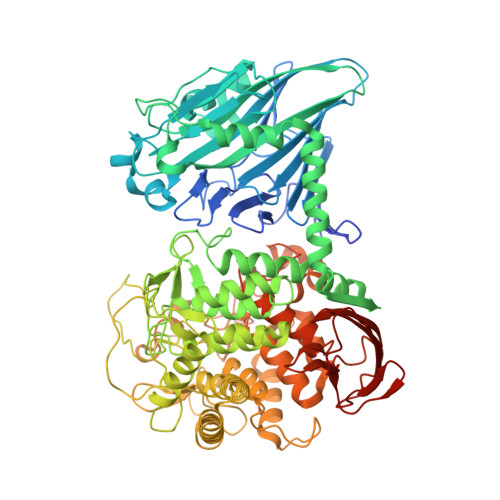 | |
UniProt | |||||
Find proteins for O66264 (Cellulomonas gilvus) Explore O66264 Go to UniProtKB: O66264 | |||||
Entity Groups | |||||
| Sequence Clusters | 30% Identity50% Identity70% Identity90% Identity95% Identity100% Identity | ||||
| UniProt Group | O66264 | ||||
Sequence AnnotationsExpand | |||||
| |||||
| Ligands 4 Unique | |||||
|---|---|---|---|---|---|
| ID | Chains | Name / Formula / InChI Key | 2D Diagram | 3D Interactions | |
| EPE Query on EPE | F [auth A] | 4-(2-HYDROXYETHYL)-1-PIPERAZINE ETHANESULFONIC ACID C8 H18 N2 O4 S JKMHFZQWWAIEOD-UHFFFAOYSA-N |  | ||
| BGC Query on BGC | C [auth A], I [auth B] | beta-D-glucopyranose C6 H12 O6 WQZGKKKJIJFFOK-VFUOTHLCSA-N |  | ||
| IFM Query on IFM | E [auth A], K [auth B] | 5-HYDROXYMETHYL-3,4-DIHYDROXYPIPERIDINE C6 H13 N O3 QPYJXFZUIJOGNX-HSUXUTPPSA-N |  | ||
| SO4 Query on SO4 | D [auth A], G [auth A], H [auth A], J [auth B], L [auth B] | SULFATE ION O4 S QAOWNCQODCNURD-UHFFFAOYSA-L |  | ||
| Length ( Å ) | Angle ( ˚ ) |
|---|---|
| a = 84.71 | α = 90 |
| b = 98.254 | β = 102.73 |
| c = 104.45 | γ = 90 |
| Software Name | Purpose |
|---|---|
| HKL-2000 | data collection |
| REFMAC | refinement |
| HKL-2000 | data reduction |
| HKL-2000 | data scaling |
RCSB PDB (citation) is hosted by
RCSB PDB is a member of the

