3QCU

Crystal structure of the LT3015 antibody Fab fragment in complex with lysophosphatidic acid (14:0)


Experimental Data Snapshot

  • Method: X-RAY DIFFRACTION
  • Resolution: 1.98 Å
  • R-Value Free: 0.252 
  • R-Value Work: 0.222 
  • R-Value Observed: 0.223 

wwPDB Validation   3D Report Full Report


Ligand Structure Quality Assessment 


This is version 1.4 of the entry. See complete history


Literature

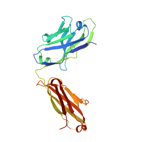
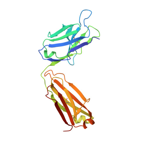
Biochemical and structural characterization of lysophosphatidic Acid binding by a humanized monoclonal antibody.

Fleming, J.K.Wojciak, J.M.Campbell, M.A.Huxford, T.

(2011) J Mol Biol 408: 462-476

  • DOI: https://doi.org/10.1016/j.jmb.2011.02.061
  • Primary Citation of Related Structures:  
    3QCT, 3QCU, 3QCV

  • PubMed Abstract: 

    Lysophosphatidic acid (LPA) is a common product of glycerophospholipid metabolism and an important mediator of signal transduction. Aberrantly high LPA concentrations accompany multiple disease states. One potential approach for treatment of these diseases, therefore, is the therapeutic application of antibodies that recognize and bind LPA as their antigen. We have determined the X-ray crystal structure of an anti-LPA antibody (LT3015) Fab fragment in its antigen-free form to 2.15 Å resolution and in complex with two LPA isotypes (14:0 and 18:2) to resolutions of 1.98 and 2.51 Å, respectively. The variable CDR (complementarity-determining region) loops at the antigen binding site adopt nearly identical conformations in the free and antigen-bound crystal structures. The crystallographic models reveal that the LT3015 antibody employs both heavy- and light-chain CDR loops to create a network of eight hydrogen bonds with the glycerophosphate head group of its LPA antigen. The head group is almost completely excluded from contact with solvent, while the hydrocarbon tail is partially solvent-exposed. In general, mutation of amino acid residues at the antigen binding site disrupts LPA binding. However, the introduction of particular mutations chosen strategically on the basis of the structures can positively influence LPA binding affinity. Finally, these structures elucidate the exquisite specificity demonstrated by an anti-lipid antibody for binding a structurally simple and seemingly unconstrained target molecule.


  • Organizational Affiliation

    Structural Biochemistry Laboratory, Department of Chemistry and Biochemistry, San Diego State University, 5500 Campanile Drive, San Diego, CA 92182-1030, USA.


Macromolecules
Find similar proteins by:  (by identity cutoff)  |  3D Structure
Entity ID: 1
MoleculeChains Sequence LengthOrganismDetailsImage
LT3015 antibody Fab fragment, heavy chainA [auth H],
C [auth I]
223Homo sapiensMutation(s): 0 
Gene Names: DKFZp686P15220
UniProt
Find proteins for Q6N089 (Homo sapiens)
Explore Q6N089 
Go to UniProtKB:  Q6N089
Entity Groups  
Sequence Clusters30% Identity50% Identity70% Identity90% Identity95% Identity100% Identity
UniProt GroupQ6N089
Sequence Annotations
Expand
  • Reference Sequence
Find similar proteins by:  (by identity cutoff)  |  3D Structure
Entity ID: 2
MoleculeChains Sequence LengthOrganismDetailsImage
LT3015 antibody Fab fragment, light chainB [auth L],
D [auth M]
218Homo sapiensMutation(s): 0 
Gene Names: IGKC
UniProt & NIH Common Fund Data Resources
Find proteins for P01834 (Homo sapiens)
Explore P01834 
Go to UniProtKB:  P01834
PHAROS:  P01834
Entity Groups  
Sequence Clusters30% Identity50% Identity70% Identity90% Identity95% Identity100% Identity
UniProt GroupP01834
Sequence Annotations
Expand
  • Reference Sequence
Small Molecules
Ligands 1 Unique
IDChains Name / Formula / InChI Key2D Diagram3D Interactions
NKN
Query on NKN

Download Ideal Coordinates CCD File 
E [auth H],
F [auth I]
(2R)-2-hydroxy-3-(phosphonooxy)propyl tetradecanoate
C17 H35 O7 P
FAZBDRGXCKPVJU-MRXNPFEDSA-N
Experimental Data & Validation

Experimental Data

  • Method: X-RAY DIFFRACTION
  • Resolution: 1.98 Å
  • R-Value Free: 0.252 
  • R-Value Work: 0.222 
  • R-Value Observed: 0.223 
  • Space Group: C 2 2 21
Unit Cell:
Length ( Å )Angle ( ˚ )
a = 86.526α = 90
b = 184.351β = 90
c = 130γ = 90
Software Package:
Software NamePurpose
DENZOdata reduction
SCALEPACKdata scaling
PHASERphasing
REFMACrefinement
PDB_EXTRACTdata extraction
HKL-2000data collection
HKL-2000data reduction
HKL-2000data scaling

Structure Validation

View Full Validation Report


Ligand Structure Quality Assessment 


Entry History 

Deposition Data

Revision History  (Full details and data files)

  • Version 1.0: 2011-03-30
    Type: Initial release
  • Version 1.1: 2011-07-13
    Changes: Version format compliance
  • Version 1.2: 2017-07-26
    Changes: Refinement description, Source and taxonomy
  • Version 1.3: 2017-11-08
    Changes: Refinement description
  • Version 1.4: 2023-09-13
    Changes: Data collection, Database references, Derived calculations, Refinement description, Structure summary