3QBT

Crystal structure of OCRL1 540-678 in complex with Rab8a:GppNHp


Experimental Data Snapshot

  • Method: X-RAY DIFFRACTION
  • Resolution: 2.00 Å
  • R-Value Free: 0.241 
  • R-Value Work: 0.208 
  • R-Value Observed: 0.210 

wwPDB Validation   3D Report Full Report


Ligand Structure Quality Assessment 


This is version 1.3 of the entry. See complete history


Literature

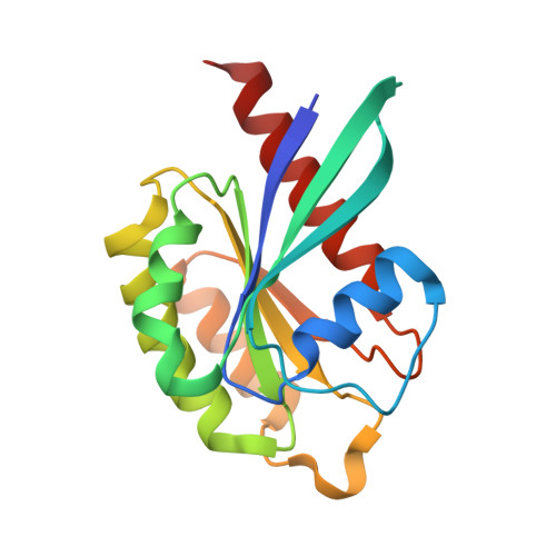
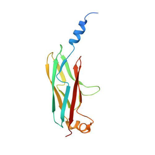
A structural basis for Lowe syndrome caused by mutations in the Rab-binding domain of OCRL1.

Hou, X.Hagemann, N.Schoebel, S.Blankenfeldt, W.Goody, R.S.Erdmann, K.S.Itzen, A.

(2011) EMBO J 30: 1659-1670

  • DOI: https://doi.org/10.1038/emboj.2011.60
  • Primary Citation of Related Structures:  
    3QBT

  • PubMed Abstract: 

    The oculocerebrorenal syndrome of Lowe (OCRL), also called Lowe syndrome, is characterized by defects of the nervous system, the eye and the kidney. Lowe syndrome is a monogenetic X-linked disease caused by mutations of the inositol-5-phosphatase OCRL1. OCRL1 is a membrane-bound protein recruited to membranes via interaction with a variety of Rab proteins. The structural and kinetic basis of OCRL1 for the recognition of several Rab proteins is unknown. In this study, we report the crystal structure of the Rab-binding domain (RBD) of OCRL1 in complex with Rab8a and the kinetic binding analysis of OCRL1 with several Rab GTPases (Rab1b, Rab5a, Rab6a and Rab8a). In contrast to other effectors that bind their respective Rab predominantly via α-helical structure elements, the Rab-binding interface of OCRL1 consists mainly of the IgG-like β-strand structure of the ASPM-SPD-2-Hydin domain as well as one α-helix. Our results give a deeper structural understanding of disease-causing mutations of OCRL1 affecting Rab binding.


  • Organizational Affiliation

    Department of Physical Biochemistry, Max-Planck-Institute of Molecular Physiology, Dortmund, Germany.


Macromolecules
Find similar proteins by:  (by identity cutoff)  |  3D Structure
Entity ID: 1
MoleculeChains Sequence LengthOrganismDetailsImage
Ras-related protein Rab-8AA,
B [auth C],
C [auth E],
D [auth G]
174Homo sapiensMutation(s): 0 
Gene Names: MELRAB8RAB8A
UniProt & NIH Common Fund Data Resources
Find proteins for P61006 (Homo sapiens)
Explore P61006 
Go to UniProtKB:  P61006
PHAROS:  P61006
GTEx:  ENSG00000167461 
Entity Groups  
Sequence Clusters30% Identity50% Identity70% Identity90% Identity95% Identity100% Identity
UniProt GroupP61006
Sequence Annotations
Expand
  • Reference Sequence
Find similar proteins by:  (by identity cutoff)  |  3D Structure
Entity ID: 2
MoleculeChains Sequence LengthOrganismDetailsImage
Inositol polyphosphate 5-phosphatase OCRL-1E [auth B],
F [auth D],
G [auth F],
H
140Homo sapiensMutation(s): 0 
Gene Names: INPP5FOCRLOCRL1
EC: 3.1.3.36
UniProt & NIH Common Fund Data Resources
Find proteins for Q01968 (Homo sapiens)
Explore Q01968 
Go to UniProtKB:  Q01968
PHAROS:  Q01968
GTEx:  ENSG00000122126 
Entity Groups  
Sequence Clusters30% Identity50% Identity70% Identity90% Identity95% Identity100% Identity
UniProt GroupQ01968
Sequence Annotations
Expand
  • Reference Sequence
Small Molecules
Ligands 3 Unique
IDChains Name / Formula / InChI Key2D Diagram3D Interactions
GNP
Query on GNP

Download Ideal Coordinates CCD File 
J [auth A],
L [auth C],
N [auth E],
P [auth G]
PHOSPHOAMINOPHOSPHONIC ACID-GUANYLATE ESTER
C10 H17 N6 O13 P3
UQABYHGXWYXDTK-UUOKFMHZSA-N
SO4
Query on SO4

Download Ideal Coordinates CCD File 
Q [auth B],
R [auth D]
SULFATE ION
O4 S
QAOWNCQODCNURD-UHFFFAOYSA-L
MG
Query on MG

Download Ideal Coordinates CCD File 
I [auth A],
K [auth C],
M [auth E],
O [auth G]
MAGNESIUM ION
Mg
JLVVSXFLKOJNIY-UHFFFAOYSA-N
Experimental Data & Validation

Experimental Data

  • Method: X-RAY DIFFRACTION
  • Resolution: 2.00 Å
  • R-Value Free: 0.241 
  • R-Value Work: 0.208 
  • R-Value Observed: 0.210 
  • Space Group: C 1 2 1
Unit Cell:
Length ( Å )Angle ( ˚ )
a = 157.147α = 90
b = 55.34β = 91.93
c = 173.836γ = 90
Software Package:
Software NamePurpose
REFMACrefinement
PDB_EXTRACTdata extraction
XSCALEdata scaling

Structure Validation

View Full Validation Report


Ligand Structure Quality Assessment 


Entry History 

Deposition Data

Revision History  (Full details and data files)

  • Version 1.0: 2011-03-23
    Type: Initial release
  • Version 1.1: 2011-07-13
    Changes: Version format compliance
  • Version 1.2: 2012-02-29
    Changes: Database references
  • Version 1.3: 2023-09-13
    Changes: Data collection, Database references, Derived calculations, Refinement description