3PY3

Crystal structure of phosphorylated p38alpha MAP kinase


Experimental Data Snapshot

  • Method: X-RAY DIFFRACTION
  • Resolution: 2.10 Å
  • R-Value Free: 0.240 
  • R-Value Work: 0.184 
  • R-Value Observed: 0.187 

wwPDB Validation   3D Report Full Report


This is version 1.3 of the entry. See complete history


Literature

Mitogen-activated Protein Kinase (MAPK) Phosphatase 3-mediated Cross-talk between MAPKs ERK2 and p38{alpha}.

Zhang, Y.Y.Wu, J.W.Wang, Z.X.

(2011) J Biol Chem 286: 16150-16162

  • DOI: https://doi.org/10.1074/jbc.M110.203786
  • Primary Citation of Related Structures:  
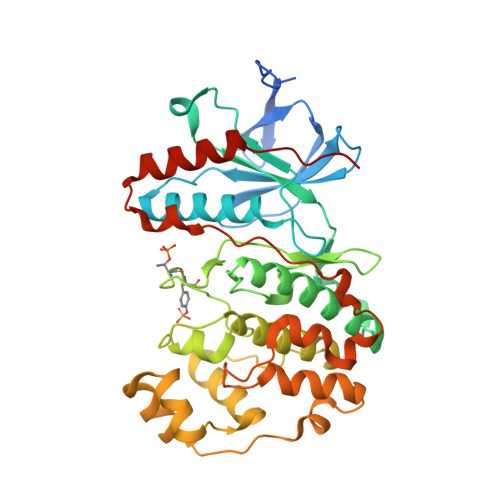
    3PY3

  • PubMed Abstract: 

    MAPK phosphatase 3 (MKP3) is highly specific for ERK1/2 inactivation via dephosphorylation of both phosphotyrosine and phosphothreonine critical for enzymatic activation. Here, we show that MKP3 is able to effectively dephosphorylate the phosphotyrosine, but not phosphothreonine, in the activation loop of p38α in vitro and in intact cells. The catalytic constant of the MKP3 reaction for p38α is comparable with that for ERK2. Remarkably, MKP3, ERK2, and phosphorylated p38α can form a stable ternary complex in solution, and the phosphatase activity of MKP3 toward p38α substrate is allosterically regulated by ERK2-MKP3 interaction. This suggests that MKP3 not only controls the activities of ERK2 and p38α but also mediates cross-talk between these two MAPK pathways. The crystal structure of bisphosphorylated p38α has been determined at 2.1 Å resolution. Comparisons between the phosphorylated MAPK structures reveal the molecular basis of MKP3 substrate specificity.


  • Organizational Affiliation

    Ministry of Education Key Laboratory of Bioinformatics, School of Life Sciences, Tsinghua University, Beijing, China.


Macromolecules
Find similar proteins by:  (by identity cutoff)  |  3D Structure
Entity ID: 1
MoleculeChains Sequence LengthOrganismDetailsImage
Mitogen-activated protein kinase 14380Mus musculusMutation(s): 0 
Gene Names: Mapk14Crk1Csbp1Csbp2
EC: 2.7.11.24
UniProt
Find proteins for P47811 (Mus musculus)
Explore P47811 
Go to UniProtKB:  P47811
Entity Groups  
Sequence Clusters30% Identity50% Identity70% Identity90% Identity95% Identity100% Identity
UniProt GroupP47811
Sequence Annotations
Expand
  • Reference Sequence
Small Molecules
Modified Residues  2 Unique
IDChains TypeFormula2D DiagramParent
PTR
Query on PTR
A
L-PEPTIDE LINKINGC9 H12 N O6 PTYR
TPO
Query on TPO
A
L-PEPTIDE LINKINGC4 H10 N O6 PTHR
Experimental Data & Validation

Experimental Data

  • Method: X-RAY DIFFRACTION
  • Resolution: 2.10 Å
  • R-Value Free: 0.240 
  • R-Value Work: 0.184 
  • R-Value Observed: 0.187 
  • Space Group: P 21 21 21
Unit Cell:
Length ( Å )Angle ( ˚ )
a = 47.272α = 90
b = 52.284β = 90
c = 168.132γ = 90
Software Package:
Software NamePurpose
HKL-2000data collection
PHASERphasing
PHENIXrefinement
HKL-2000data reduction
HKL-2000data scaling

Structure Validation

View Full Validation Report


Entry History 

Deposition Data

Revision History  (Full details and data files)

  • Version 1.0: 2011-03-16
    Type: Initial release
  • Version 1.1: 2011-07-13
    Changes: Version format compliance
  • Version 1.2: 2023-11-01
    Changes: Data collection, Database references, Derived calculations, Refinement description
  • Version 1.3: 2023-12-06
    Changes: Data collection