3POT

Structural analysis of a Ni(III)-methyl species in methyl-coenzyme M reductase from Methanothermobacter marburgensis


Experimental Data Snapshot

  • Method: X-RAY DIFFRACTION
  • Resolution: 1.20 Å
  • R-Value Free: 0.156 
  • R-Value Work: 0.132 
  • R-Value Observed: 0.133 

wwPDB Validation   3D Report Full Report


Ligand Structure Quality Assessment 


This is version 1.4 of the entry. See complete history


Literature

Structural Analysis of a Ni-Methyl Species in Methyl-Coenzyme M Reductase from Methanothermobacter marburgensis.

Cedervall, P.E.Dey, M.Li, X.Sarangi, R.Hedman, B.Ragsdale, S.W.Wilmot, C.M.

(2011) J Am Chem Soc 133: 5626-5628

  • DOI: https://doi.org/10.1021/ja110492p
  • Primary Citation of Related Structures:  
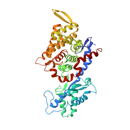
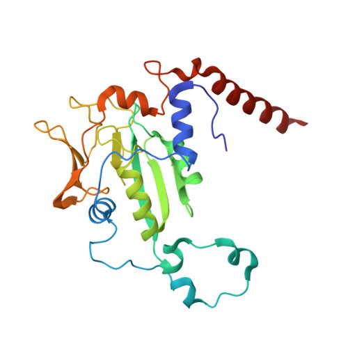
    3POT

  • PubMed Abstract: 

    We present the 1.2 Å resolution X-ray crystal structure of a Ni-methyl species that is a proposed catalytic intermediate in methyl-coenzyme M reductase (MCR), the enzyme that catalyzes the biological formation of methane. The methyl group is situated 2.1 Å proximal of the Ni atom of the MCR coenzyme F(430). A rearrangement of the substrate channel has been posited to bring together substrate species, but Ni(III)-methyl formation alone does not lead to any observable structural changes in the channel.


  • Organizational Affiliation

    Department of Biochemistry, Molecular Biology and Biophysics, University of Minnesota, Minneapolis, Minnesota 55455, United States.


Macromolecules
Find similar proteins by:  (by identity cutoff)  |  3D Structure
Entity ID: 1
MoleculeChains Sequence LengthOrganismDetailsImage
Methyl-coenzyme M reductase I subunit alpha
A, D
550Methanothermobacter marburgensisMutation(s): 0 
EC: 2.8.4.1
UniProt
Find proteins for P11558 (Methanothermobacter marburgensis (strain ATCC BAA-927 / DSM 2133 / JCM 14651 / NBRC 100331 / OCM 82 / Marburg))
Explore P11558 
Go to UniProtKB:  P11558
Entity Groups  
Sequence Clusters30% Identity50% Identity70% Identity90% Identity95% Identity100% Identity
UniProt GroupP11558
Sequence Annotations
Expand
  • Reference Sequence
Find similar proteins by:  (by identity cutoff)  |  3D Structure
Entity ID: 2
MoleculeChains Sequence LengthOrganismDetailsImage
Methyl-coenzyme M reductase I subunit beta
B, E
443Methanothermobacter marburgensisMutation(s): 0 
EC: 2.8.4.1
UniProt
Find proteins for P11560 (Methanothermobacter marburgensis (strain ATCC BAA-927 / DSM 2133 / JCM 14651 / NBRC 100331 / OCM 82 / Marburg))
Explore P11560 
Go to UniProtKB:  P11560
Entity Groups  
Sequence Clusters30% Identity50% Identity70% Identity90% Identity95% Identity100% Identity
UniProt GroupP11560
Sequence Annotations
Expand
  • Reference Sequence
Find similar proteins by:  (by identity cutoff)  |  3D Structure
Entity ID: 3
MoleculeChains Sequence LengthOrganismDetailsImage
Methyl-coenzyme M reductase I subunit gamma
C, F
249Methanothermobacter marburgensisMutation(s): 0 
EC: 2.8.4.1
UniProt
Find proteins for P11562 (Methanothermobacter marburgensis (strain ATCC BAA-927 / DSM 2133 / JCM 14651 / NBRC 100331 / OCM 82 / Marburg))
Explore P11562 
Go to UniProtKB:  P11562
Entity Groups  
Sequence Clusters30% Identity50% Identity70% Identity90% Identity95% Identity100% Identity
UniProt GroupP11562
Sequence Annotations
Expand
  • Reference Sequence
Small Molecules
Ligands 8 Unique
IDChains Name / Formula / InChI Key2D Diagram3D Interactions
F43
Query on F43

Download Ideal Coordinates CCD File 
K [auth A],
V [auth D]
FACTOR 430
C42 H51 N6 Ni O13
XLFIRMYGVLUNOY-SXMZNAGASA-M
TP7
Query on TP7

Download Ideal Coordinates CCD File 
N [auth A],
W [auth D]
Coenzyme B
C11 H22 N O7 P S
JBJSVEVEEGOEBZ-SCZZXKLOSA-N
TXZ
Query on TXZ

Download Ideal Coordinates CCD File 
M [auth A],
X [auth D]
O-phosphono-N-(6-sulfanylhexanoyl)-L-threonine
C10 H20 N O7 P S
PRNNQVAYHYUTOD-APPZFPTMSA-N
COM
Query on COM

Download Ideal Coordinates CCD File 
L [auth A],
Y [auth D]
1-THIOETHANESULFONIC ACID
C2 H6 O3 S2
ZNEWHQLOPFWXOF-UHFFFAOYSA-N
06C
Query on 06C

Download Ideal Coordinates CCD File 
O [auth A],
Z [auth D]
Iodomethane
C H3 I
INQOMBQAUSQDDS-UHFFFAOYSA-N
EDO
Query on EDO

Download Ideal Coordinates CCD File 
AA [auth D]1,2-ETHANEDIOL
C2 H6 O2
LYCAIKOWRPUZTN-UHFFFAOYSA-N
K
Query on K

Download Ideal Coordinates CCD File 
P [auth A]POTASSIUM ION
K
NPYPAHLBTDXSSS-UHFFFAOYSA-N
MG
Query on MG

Download Ideal Coordinates CCD File 
BA [auth E]
CA [auth E]
DA [auth F]
G [auth A]
H [auth A]
BA [auth E],
CA [auth E],
DA [auth F],
G [auth A],
H [auth A],
I [auth A],
J [auth A],
Q [auth B],
R [auth B],
S [auth C],
T [auth C],
U [auth D]
MAGNESIUM ION
Mg
JLVVSXFLKOJNIY-UHFFFAOYSA-N
Modified Residues  5 Unique
IDChains TypeFormula2D DiagramParent
AGM
Query on AGM
A, D
L-PEPTIDE LINKINGC7 H17 N4 O2ARG
GL3
Query on GL3
A, D
L-PEPTIDE LINKINGC2 H5 N O SGLY
MGN
Query on MGN
A, D
L-PEPTIDE LINKINGC6 H12 N2 O3GLN
MHS
Query on MHS
A, D
L-PEPTIDE LINKINGC7 H11 N3 O2HIS
SMC
Query on SMC
A, D
L-PEPTIDE LINKINGC4 H9 N O2 SCYS
Experimental Data & Validation

Experimental Data

  • Method: X-RAY DIFFRACTION
  • Resolution: 1.20 Å
  • R-Value Free: 0.156 
  • R-Value Work: 0.132 
  • R-Value Observed: 0.133 
  • Space Group: P 1 21 1
Unit Cell:
Length ( Å )Angle ( ˚ )
a = 81.886α = 90
b = 118.165β = 91.94
c = 122.388γ = 90
Software Package:
Software NamePurpose
REFMACrefinement
HKL-2000data reduction
HKL-2000data scaling
REFMACphasing

Structure Validation

View Full Validation Report


Ligand Structure Quality Assessment 


Entry History 

Deposition Data

Revision History  (Full details and data files)

  • Version 1.0: 2011-04-13
    Type: Initial release
  • Version 1.1: 2011-07-13
    Changes: Version format compliance
  • Version 1.2: 2015-05-20
    Changes: Non-polymer description
  • Version 1.3: 2017-03-29
    Changes: Non-polymer description
  • Version 1.4: 2023-09-06
    Changes: Data collection, Database references, Derived calculations, Refinement description