


Lipid binding protein
Lu, Y.Z., Zhao, X.To be published.
Experimental Data Snapshot
Entity ID: 1 | |||||
|---|---|---|---|---|---|
| Molecule | Chains | Sequence Length | Organism | Details | Image |
| Putative uncharacterized protein | 320 | Streptococcus mutans UA159 | Mutation(s): 1 Gene Names: SMU_165 | 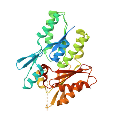 | |
UniProt | |||||
Find proteins for Q8DWA2 (Streptococcus mutans serotype c (strain ATCC 700610 / UA159)) Explore Q8DWA2 Go to UniProtKB: Q8DWA2 | |||||
Entity Groups | |||||
| Sequence Clusters | 30% Identity50% Identity70% Identity90% Identity95% Identity100% Identity | ||||
| UniProt Group | Q8DWA2 | ||||
Sequence AnnotationsExpand | |||||
| |||||
| Ligands 1 Unique | |||||
|---|---|---|---|---|---|
| ID | Chains | Name / Formula / InChI Key | 2D Diagram | 3D Interactions | |
| PLM Query on PLM | B [auth A] | PALMITIC ACID C16 H32 O2 IPCSVZSSVZVIGE-UHFFFAOYSA-N |  | ||
| Length ( Å ) | Angle ( ˚ ) |
|---|---|
| a = 42.304 | α = 90 |
| b = 42.304 | β = 90 |
| c = 151.314 | γ = 90 |
| Software Name | Purpose |
|---|---|
| CrysalisPro | data collection |
| MOLREP | phasing |
| REFMAC | refinement |
| CrysalisPro | data reduction |
| SCALA | data scaling |
RCSB PDB (citation) is hosted by
RCSB PDB is a member of the

