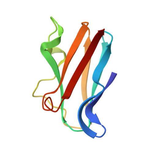
The crystal structure of mercury-substituted poplar plastocyanin at 1.9-A resolution.
Church, W.B., Guss, J.M., Potter, J.J., Freeman, H.C.(1986) J Biol Chem 261: 234-237
- PubMed: 3941073
- DOI: https://doi.org/10.2210/pdb3pcy/pdb
- Primary Citation of Related Structures:
3PCY - PubMed Abstract:
The crystal structure of Hg(II)-plastocyanin has been determined and refined at a resolution of 1.9 A. The crystals were prepared by soaking crystals of Cu(II)-plastocyanin from poplar leaves (Populus nigra var. italica) in a solution of a mercuric salt. Replacement of the Cu(II) atom in plastocyanin by Hg(II) causes only minor changes in the geometry of the metal site, and there are few significant changes elsewhere in the molecule. It is concluded that, as in the case of the native protein, the geometry of the metal site is determined by the polypeptide. The weak metal-S(methionine) bond found in Cu(II)-plastocyanin remains weak in Hg(II)-plastocyanin. The "flip" of a proline side chain close to the metal site from a C gamma-exo conformation in Cu(II)-plastocyanin to a C gamma-endo conformation in Hg(II)-plastocyanin suggests that this region of the molecule is particularly flexible. Crystallographic evidence for the close similarity of the Hg(II)- and Cu(II)-plastocyanin structures was originally obtained from electron density difference maps at 2.5-A resolution. The refinement of the structure was begun with a set of atomic coordinates taken from the structure of Cu(II)-plastocyanin. A Hg(II) atom was substituted for the Cu(II) atom, and the side chains of 6 residues in the vicinity of the metal site were omitted. Three series of stereochemically restrained least-squares refinement calculations were interspersed with two stages of model adjustment followed by phase extension. Fifty-nine water molecules were located. The final structure has a crystallographic residual R = 0.16.


