3P76

X-ray crystal structure of Aquifex aeolicus LpxC complexed SCH1379777


Experimental Data Snapshot

  • Method: X-RAY DIFFRACTION
  • Resolution: 1.93 Å
  • R-Value Free: 0.185 
  • R-Value Work: 0.168 
  • R-Value Observed: 0.169 

wwPDB Validation   3D Report Full Report


Ligand Structure Quality Assessment 


This is version 1.2 of the entry. See complete history


Literature

Design and synthesis of potent Gram-negative specific LpxC inhibitors.

Faruk Mansoor, U.Vitharana, D.Reddy, P.A.Daubaras, D.L.McNicholas, P.Orth, P.Black, T.Arshad Siddiqui, M.

(2011) Bioorg Med Chem Lett 21: 1155-1161

  • DOI: https://doi.org/10.1016/j.bmcl.2010.12.111
  • Primary Citation of Related Structures:  
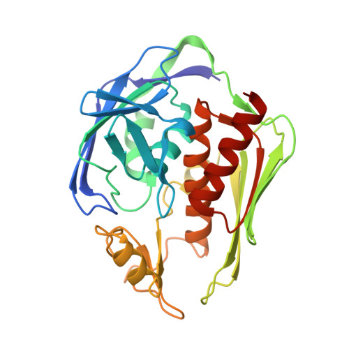
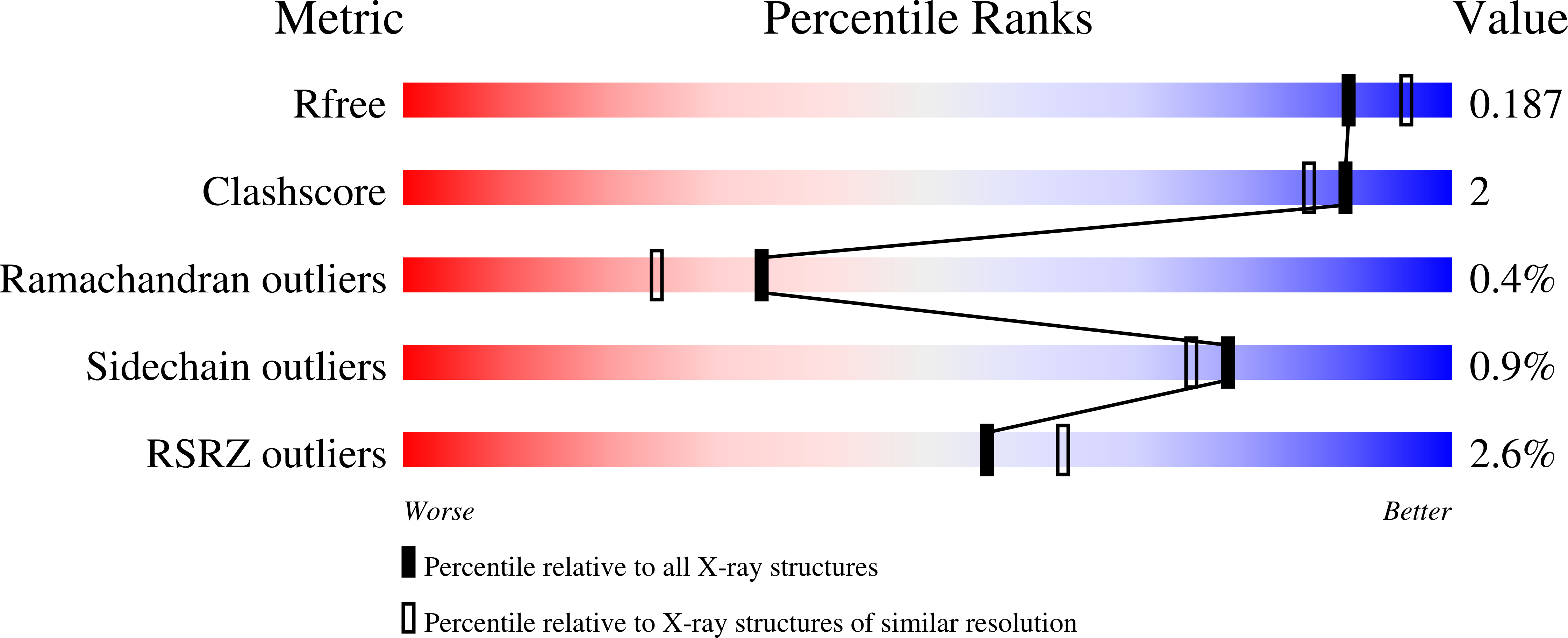
    3P76

  • PubMed Abstract: 

    Antibiotic resistant hospital acquired infections are on the rise, creating an urgent need for novel bactericidal drugs. Enzymes involved in lipopolysaccharide (LPS) biosynthesis are attractive antibacterial targets since LPS is the major structural component of the outer membrane of Gram-negative bacteria. Lipid A is an essential hydrophobic anchor of LPS and the first committed step in lipid A biosynthesis is catalyzed by a unique zinc dependent metalloamidase, UDP-3-O-(R-3-hydroxymyristoyl)-N-acetylglucosamine deacetylase (LpxC). LpxC is an attractive Gram-negative only target that has been chemically validated by potent bactericidal hydroxamate inhibitors that work by coordination of the enzyme's catalytic zinc ion. An exploratory chemistry effort focused on expanding the SAR around hydroxamic acid zinc-binding 'warheads' lead to the identification of novel compounds with enzyme potency and antibacterial activity similar to CHIR-090.


  • Organizational Affiliation

    Department of Chemistry, Merck Research Laboratories, 320 Bent Street, Cambridge, MA 02141, USA. umar.faruk.mansoor@merck.com


Macromolecules
Find similar proteins by:  (by identity cutoff)  |  3D Structure
Entity ID: 1
MoleculeChains Sequence LengthOrganismDetailsImage
UDP-3-O-[3-hydroxymyristoyl] N-acetylglucosamine deacetylase271Aquifex aeolicusMutation(s): 0 
EC: 3.5.1
UniProt
Find proteins for O67648 (Aquifex aeolicus (strain VF5))
Explore O67648 
Go to UniProtKB:  O67648
Entity Groups  
Sequence Clusters30% Identity50% Identity70% Identity90% Identity95% Identity100% Identity
UniProt GroupO67648
Sequence Annotations
Expand
  • Reference Sequence
Small Molecules
Binding Affinity Annotations 
IDSourceBinding Affinity
P76 PDBBind:  3P76 IC50: 2.1 (nM) from 1 assay(s)
Experimental Data & Validation

Experimental Data

  • Method: X-RAY DIFFRACTION
  • Resolution: 1.93 Å
  • R-Value Free: 0.185 
  • R-Value Work: 0.168 
  • R-Value Observed: 0.169 
  • Space Group: P 61
Unit Cell:
Length ( Å )Angle ( ˚ )
a = 65.592α = 90
b = 65.592β = 90
c = 133.588γ = 120
Software Package:
Software NamePurpose
DENZOdata reduction
SCALEPACKdata scaling
PDB_EXTRACTdata extraction
CrystalCleardata collection
AMoREphasing
BUSTERrefinement

Structure Validation

View Full Validation Report


Ligand Structure Quality Assessment 


Entry History 

Deposition Data

  • Released Date: 2011-02-09 
  • Deposition Author(s): Orth, P.

Revision History  (Full details and data files)

  • Version 1.0: 2011-02-09
    Type: Initial release
  • Version 1.1: 2011-07-13
    Changes: Version format compliance
  • Version 1.2: 2023-09-06
    Changes: Data collection, Database references, Derived calculations, Refinement description